| disease | Odontoid Process Developmental Deformity |
The odontoid process is a crucial bony connecting structure in the upper cervical joint. It maintains the stability of the atlantoaxial joint by being secured within a specific anatomical range by the transverse ligament of the atlas. Hypoplasia of the odontoid process and the transverse ligament are the primary congenital factors leading to atlantoaxial instability. Currently, it has been found that such deformities are not uncommon, accounting for approximately 4/5 of occipitocervical deformities.
bubble_chart Etiology
The exact cause of odontoid process developmental abnormalities is not entirely clear. The odontoid process originates from the mesenchymal tissue of the first cervical vertebra during the embryonic period. During the development of the odontoid process, there are initially two ossification centers that appear around the fifth month of embryonic development and soon fuse into a single ossification center. The epiphyseal plate of this ossification center is located between the odontoid process and the body of the axis vertebra. Under normal circumstances, this epiphyseal plate completely fuses by around the age of 5, integrating the odontoid process with the axis vertebra. During this developmental process, certain congenital factors can lead to the absence or hypoplasia of the odontoid process. Additionally, the persistence of mesenchymal tissue between the odontoid process and the body of the axis vertebra without undergoing chondrification and ossification can result in odontoid process malformation. Furthermore, acquired trauma or infection can affect the blood supply to the tip of the odontoid process, leading to its hypoplasia.
bubble_chart Pathological ChangesThe atlantoaxial joint consists of three joints: the bilateral lateral atlantoaxial joints and the median atlantoaxial joint in the middle. The lateral atlantoaxial joint is formed by the inferior articular surface of the atlas and the superior articular surface of the axis, with the joint plane being horizontal. The median atlantoaxial joint is further composed of two joints: one between the anterior surface of the odontoid process and the odontoid fossa of the atlas, and the other between the posterior surface of the odontoid process and the ligament of the atlas. Below the inner edge of the superior articular surfaces of the axis on both sides, there is a small tubercle where the transverse ligament of the atlas attaches. This ligament is very strong and is the main structure preventing anterior displacement of the atlas (Figure 1). It is evident that the odontoid process and the transverse ligament are crucial factors in stabilizing the atlantoaxial joint. Additionally, the alar ligament and the apical ligament of the odontoid process attach to the anterior margin of the foramen magnum and the medial surface of the occipital condyle, respectively, playing a role in maintaining the stability of the atlantoaxial joint.

Figure 1 Cross-sectional view of the odontoid process
Odontoid process abnormalities result in the loss of normal physiological control of the atlantoaxial joint, inevitably leading to instability of the atlantoaxial joint. In cases of odontoid process absence or hypoplasia, the interlocking relationship between the transverse ligament of the atlas and the odontoid process is lost, causing anterior or rotational dislocation of the atlas and resulting in spinal cord compression. In cases where the tip and base of the odontoid process fail to fuse, the odontoid process can move with the atlas, leading to laxity of the transverse ligament. Over time, other ligamentous structures such as the alar ligament and the apical ligament also become lax, ultimately resulting in atlantoaxial dislocation and spinal cord compression (Figure 2). Furthermore, due to atlantoaxial joint instability, the lateral atlantoaxial joints undergo degeneration from long-term friction and stimulation, and the resulting bone hyperplasia can exacerbate spinal cord compression. The atlanto-occipital membrane thickens into a band-like structure due to friction and stimulation, similarly increasing spinal cord compression.

Figure 2 Anterior dislocation of the atlas and spinal cord compression
bubble_chart Clinical Manifestations
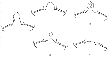
Odontoid process malformations can be divided into three types: odontoid hypoplasia, os odontoideum, and odontoid aplasia, with odontoid aplasia being relatively rare (Figure 3). Sometimes, the os odontoideum can be confused with an odontoid fracture nonunion. The distinction lies in the fact that the os odontoideum is smaller and smoother, located above the atlantoaxial joint space; whereas an odontoid fracture nonunion has a fracture line, normal development, and is mostly at the level of the atlantoaxial joint.

Figure 3 Common classification of odontoid process malformations
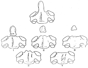
Greenberg classified odontoid process malformations into five types (Figure 4):

Figure 4 Greenberg classification of odontoid process malformations
Type I: Free os odontoideum, where the odontoid process is not fused with the axis.
Type II: Absence of the waist of the odontoid process, with a free small bone at the tip of the odontoid, separated from the base.
Type III: Hypoplasia of the base of the odontoid process, with only the tip of the odontoid remaining.
Type IV: Absence of the tip of the odontoid process.
Type V: Complete absence of the odontoid process.
Professor Jia Lianshun discovered another type of developmental disorder in recent studies, where the odontoid process is short and thick, resembling a complete odontoid process but significantly shorter than normal, with a wider base, which can be referred to as "short odontoid type malformation" (Figure 5).

Figure 5 "Short odontoid type" odontoid process malformation
The clinical manifestations of various types of odontoid process malformations are generally similar. In the early stages, due to limited activity, there may be no atlantoaxial instability or symptoms of nerve compression, but there is potential instability, with a significantly increased range of passive head movement, increased atlantoaxial movement, and X-ray showing grade I anterior displacement of the atlas. Some cases may have the malformation for life without symptoms. Most cases, with increasing age, increased cervical activity, or minor trauma, may lead to atlantoaxial joint dislocation or subluxation, resulting in clinical symptoms of spinal cord compression. The main manifestations include head and neck pain, weak neck muscles unable to support the head, weakness in both lower limbs, unsteady walking, and impaired fine motor skills of the fingers, which may progress to partial or complete spastic paralysis of the limbs, and even sudden death. Some patients may show clinical manifestations of vertebral artery insufficiency; a few patients may experience difficulty breathing and dysfunction of bowel and bladder. Signs mainly include restricted cervical movement, prominence and tenderness of the axis spinous process, tenderness of the paravertebral muscles, and straightening of the occipital cervical curve; there may be increased muscle tone in the limbs, hyperactive or exaggerated tendon reflexes, positive pathological reflexes such as Hoffmann's sign and Babinski's sign, and elicitable patellar and ankle clonus. In severe cases, high cervical spinal cord compression symptoms may occur, manifesting as difficulty breathing or respiratory paralysis. Odontoid process malformations are more common in patients with bone dysplasia, such as mucopolysaccharidosis, spondyloepiphyseal dysplasia dwarfism, etc., and may also be associated with other occipital cervical malformations such as basilar invagination or platybasia.
bubble_chart DiagnosisClinical manifestations combined with imaging examinations can provide a definitive diagnosis of congenital odontoid process malformation and dislocation of atlantoaxial vertebrae.
X-ray examination includes anteroposterior and lateral views of the cervical spine, dynamic lateral views in flexion and extension, and open-mouth anteroposterior views. If necessary, tomographic imaging during a seasonal epidemic can be performed. This allows observation of the characteristics of odontoid process malformation and the status of dislocation of atlantoaxial vertebrae, and can infer the state of spinal cord compression. The specific features on X-ray images are as follows: (1) In cases of odontoid process absence or hypoplasia, a short or absent odontoid process can be seen on lateral and open-mouth anteroposterior views of the atlantoaxial vertebrae; (2) In cases of os odontoideum, the free odontoid bone is connected to the anterior arch of the atlas and shows a significant gap with the body of the axis. Dynamic lateral views in flexion and extension can reveal the free odontoid bone moving forward with the atlas.
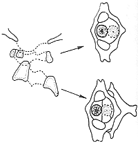
CT scan examination allows understanding of the type of odontoid process malformation and the degree of dislocation of atlantoaxial vertebrae through analysis of images on scanning layers. (1) In cases of odontoid process absence, no odontoid process appears on the corresponding scanning layers; (2) In cases of odontoid process hypoplasia, only a small odontoid shadow or punctate ossification shadow appears on the scanning layers; (3) In cases of os odontoideum, a double odontoid shadow may appear within the atlas ring, indicating that the odontoid process moves forward with the atlas (Figures 1 and 2).

Figure 1: CT slice showing two bone shadows within the atlas ring, including the free small bone of the odontoid process.

Figure 2: Different CT scan slices showing the positions of the odontoid process and the atlas.
Magnetic resonance imaging (MRI) examination can reveal the dislocation of atlantoaxial vertebrae caused by odontoid process malformation and the state of spinal cord compression. It also provides information on the relationships between bones, ligaments, dura mater, and the spinal cord, offering reliable evidence for designing treatment plans. The main MRI findings of odontoid process malformation and atlantoaxial instability include synchronous forward displacement of the anterior and posterior arch nodules of the atlas, with the free odontoid process moving forward synchronously with the atlas, and showing the state of spinal cord compression.
bubble_chart Treatment Measures
1. For congenital odontoid process malformation without neurological symptoms, active treatment measures should generally be adopted. For the elderly or younger children, neck movement should be reduced to prevent trauma, and a cervical collar should be used locally to maintain or slow down its progression. At the same time, closely monitor the condition. Once neurological compression symptoms appear, active surgical treatment should be undertaken to stabilize the atlantoaxial joint.
2. Surgical treatment should be provided for odontoid process malformation causing significant atlantoaxial instability combined with spinal cord compression. Surgical methods include: (1) simple occipitocervical fusion; (2) atlantoaxial fusion; (3) decompression and occipitocervical fusion. The author once designed posterior arch resection of the atlas and occipitocervical fusion, achieving good results. In recent years, Magerl designed the posterior atlantoaxial lateral joint screw fixation, which has the advantage of immediately obtaining a firm fixation of the atlantoaxial joint postoperatively without the need for gypsum bed fixation.
3. Congenital odontoid process malformation combined with basilar invagination, occipitalization of the atlas, or narrowing of the foramen magnum. In such cases, due to the coexistence of multiple malformations, there are multiple factors causing spinal cord compression, with the posterior edge of the foramen magnum being a significant compressing factor. Simple occipitocervical fusion cannot achieve the treatment goal. Foramen magnum enlargement and posterior arch resection of the atlas with decompression and bone graft fusion can be performed. This surgery can directly remove the compressing factor and stabilize the atlantoaxial joint.
Key points of the surgery: (1) Exposure of the occipitocervical region: Make a posterior midline incision from 2.0 cm above the occipital protuberance to the spinous process of the fourth cervical vertebra. The exposure process is divided into three steps: occipital bone, cervical vertebrae, and the occipitocervical junction. When the atlas is displaced forward, the posterior arch is deeper, so it should be first touched with a finger, followed by careful sharp dissection. The exposure range of the posterior arch is limited to 1.5 cm on each side of the posterior arch tubercle to avoid injury to the vertebral artery; (2) Foramen magnum enlargement and posterior arch resection of the atlas for decompression: First, drill a hole or use a small sharp chisel to create a hole 2.0 cm to 2.5 cm above the posterior foramen magnum, then extend towards the foramen magnum with an impact rongeur, and finally remove the posterior edge of the foramen magnum and the posterior arch of the atlas. Due to the forward displacement of the atlas, its position is deeper and closely in contact with the dura mater, so the resection should be done very carefully, ensuring full mobilization before removal. Additionally, the fibrous bands formed by long-term friction between the posterior edge of the foramen magnum and the posterior arch of the atlas with the dura mater should be longitudinally incised to fully decompress the spinal cord; (3) Bone graft fusion: After foramen magnum enlargement and decompression, create a bone groove 2.0 cm above the foramen magnum and implant the bone graft strip, with its lower end trimmed into a fishtail shape to tightly interlock with the base of the second cervical vertebra's spinous process. The deep soft tissues should be tightly sutured to firmly fix the bone graft strip; (4) Postoperative gypsum bed fixation, replaced with head-neck-thorax gypsum fixation after suture removal.
Dislocation of atlantoaxial vertebrae caused by odontoid process anomalies should be differentiated from traumatic dislocation, spontaneous dislocation, and pathological dislocation.


