| disease | Cervical Spinal Canal Stenosis |
The narrowing of the cervical spinal canal caused by bony or fibrous degeneration due to developmental or degenerative factors in the anatomical structures constituting the cervical spinal canal, leading to impaired spinal cord blood circulation and compression of the spinal cord and nerve roots, is termed cervical spinal canal stenosis. Clinically, lumbar spinal canal stenosis is the most common, followed by cervical spinal canal stenosis, while thoracic spinal canal stenosis is the least common. Spinal canal stenosis was first described in 1900 by Sachs and Fraenkel in a report on the treatment of lumbar spinal canal stenosis using two-level laminectomy. Cervical spinal canal stenosis is a concept that was gradually recognized later. In 1976, Arnold et al. classified spinal canal stenosis into congenital and acquired types. Congenital spinal canal stenosis refers to narrowing caused by developmental disorders of the vertebral arch before or after birth, with developmental spinal canal stenosis limited to vertebral arch developmental disorders being the most common, also known as idiopathic spinal canal stenosis. The primary disease cause of acquired spinal canal stenosis is degenerative changes in the spine. Cervical spinal canal stenosis is more common in middle-aged and elderly individuals, with the lower cervical spine being the most frequently affected area, particularly the C4–6 segments, and the onset is slow.
bubble_chart Etiology
Cervical spinal canal stenosis is classified into four categories based on disease cause: (1) developmental cervical spinal canal stenosis; (2) degenerative cervical spinal canal stenosis; (3) iatrogenic cervical spinal canal stenosis; (4) secondary cervical spinal canal stenosis caused by other pathologies and trauma, such as cervical spondylosis, cervical disc herniation, ossification of the posterior longitudinal ligament, cervical subcutaneous nodules, tumors, and trauma. However, these conditions belong to different categories of cervical spine disorders.
bubble_chart Pathological Changes1. Developmental cervical spinal canal stenosis
In the early stages or in the absence of external traumatic factors, symptoms may not appear. However, with degenerative changes in the spine (such as bone spurs, herniated intervertebral discs, segmental instability, etc.), or after a trauma to the head and neck, the spinal canal may further narrow, leading to a series of clinical manifestations of spinal cord compression. Due to the reduced or absent reserve space in the stenotic spinal canal, the spinal cord becomes closer to the anterior and posterior walls of the canal. Even during normal cervical flexion and extension movements, irritation or compression may occur, resulting in spinal cord damage. When secondary factors such as trauma, segmental instability, or herniation or prolapse of the nucleus pulposus are present—especially when sudden external force is applied to the head and neck—it may cause significant relative displacement of the intervertebral joints, herniation of the intervertebral disc or rupture, infolding of the ligamentum flavum into the spinal canal, and changes in the sagittal diameter of the spinal cord. These instantaneous changes inevitably alter the sagittal diameter of the spinal canal. Since developmental spinal canal stenosis already has minimal reserve space, the spinal cord or nerve roots cannot tolerate even these minor changes in internal diameter, leading to injury. Since the 1970s, developmental spinal canal stenosis has been recognized as an important disease cause of cervical spondylosis-related myelopathy. Clinical data indicate that 60–70% of patients with cervical spondylotic myelopathy have developmental cervical spinal canal stenosis.
This is the most common type of cervical spinal canal stenosis. After middle age, the cervical spine gradually undergoes degenerative changes. The timing and extent of degeneration are closely related to individual differences, occupation, labor intensity, and trauma. The cervical spine is located between the relatively fixed thoracic spine and the skull and is highly mobile, making it prone to strain after middle age. The first degenerative changes occur in the intervertebral discs, followed by the ligaments, joint capsules, and bones. Degenerative changes in the intervertebral discs lead to segmental instability, osteophyte formation at the posterior vertebral margins, thickening of the lamina, hypertrophy of the facet joints, and thickening of the ligamentum flavum, resulting in anterior compression of the spinal cord by protruding structures. The thickened ligamentum flavum may fold during cervical extension, irritating and compressing the spinal cord from behind. This reduces the effective volume of the spinal canal, greatly diminishing or even eliminating the buffer space within the canal, leading to compression of the corresponding cervical spinal cord segments. If trauma occurs at this stage, it may damage the bony or fibrous structures within the canal, rapidly manifesting as spinal cord compression, as the degenerated intervertebral discs are more prone to rupture.
3. Iatrogenic cervical spinal canal stenosis
This condition is caused by surgical intervention. The main reasons include: (1) surgical trauma and hemorrhage leading to scar tissue formation, which adheres to the dura mater and compresses the spinal cord; (2) excessive or overly extensive laminectomy without bony fusion, resulting in cervical instability and secondary traumatic or fibrotic proliferative changes; (3) bone graft protrusion into the spinal canal after anterior cervical decompression and fusion; (4) failure of cervical laminoplasty, such as hinge fracture.
These include cervical spondylosis, cervical disc herniation, ossification of the posterior longitudinal ligament (OPLL), cervical tumors, subcutaneous nodes, and trauma. However, these are independent diseases, and cervical spinal canal stenosis is only part of their pathological manifestations. Therefore, they should not be diagnosed as cervical spinal canal stenosis.
bubble_chart Clinical Manifestations
Sensory disturbances are mainly manifested as numbness, hypersensitivity, or pain in the limbs. Most patients experience these symptoms, which are often the initial symptoms. This is primarily caused by the involvement of the spinothalamic tract and other sensory nerve fiber bundles. Symptoms may occur simultaneously in all limbs or start in one limb first, but in most patients, sensory disturbances begin in the upper limbs, particularly in the arms. Trunk symptoms include sensory disturbances below the second or fourth ribs, tightness in the chest, abdomen, or pelvic region, referred to as a "band-like sensation." In severe cases, difficulty breathing may occur.
Motor disturbances often appear after sensory disturbances, manifesting as pyramidal tract signs, such as limb weakness, stiffness, and clumsiness. Most cases begin with weakness and heaviness in the lower limbs, with a sensation of stepping on cotton when walking. In severe cases, standing and walking become unsteady, leading to frequent falls, requiring support from walls or crutches. As symptoms gradually worsen, paralysis of the limbs may occur.
Bowel and bladder dysfunction usually appear later. Early symptoms include weakness in urination and defecation, with frequent urination, urgency, and constipation being common. In advanced stages, urinary retention and fecal incontinence may occur.
Signs: Neck symptoms are minimal, with no significant restriction in cervical spine movement. Mild tenderness may be present over the cervical spinous processes or adjacent muscles. Sensory disturbances in the trunk and limbs are common but irregular. The trunk may show asymmetrical sensory levels, or there may be a segmental area of sensory loss with normal sensation below the waist. Superficial reflexes, such as the abdominal reflex and cremasteric reflex, are often weakened or absent. Deep sensations, such as position sense and vibration sense, remain intact. The anal reflex is usually preserved, while tendon reflexes are often hyperactive or exaggerated. A unilateral or bilateral positive Hoffmann's sign is an important indicator of spinal cord compression above the cervical 6 level. In the lower limbs, spasticity may lead to a positive Babinski sign and positive patellar or ankle clonus. Muscle atrophy, decreased muscle strength, and increased muscle tone are observed in the limbs. Muscle atrophy appears early and is widespread, especially in patients with developmental cervical spinal canal stenosis, as the pathology involves multiple segments. Once the cervical spinal cord is affected, multiple segments are often involved, but the affected level generally does not exceed the nerve distribution of the highest stenotic segment.
bubble_chart Auxiliary Examination
Imaging Examination:
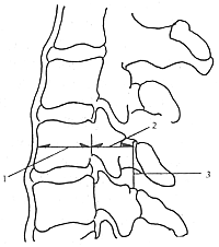
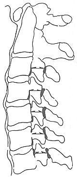
I. X-ray Plain Film Examination Developmental cervical spinal canal stenosis primarily manifests as a reduction in the sagittal diameter of the cervical spinal canal. Therefore, measuring the sagittal diameter of the spinal canal on a standard lateral radiograph is an accurate and straightforward method for diagnosis. The sagittal diameter of the spinal canal is defined as the shortest distance from the posterior edge of the vertebral body to the baseline of the spinous process. An absolute sagittal diameter of less than 12 mm indicates developmental cervical spinal canal stenosis, while a diameter of less than 10 mm signifies absolute stenosis. The ratio method is more precise because the median sagittal planes of the spinal canal and vertebral body lie on the same anatomical plane, sharing the same magnification factor, thus eliminating magnification effects (Figure 1). The normal spinal canal-to-vertebral body ratio is 1:1. A ratio of less than 0.82:1 suggests spinal canal stenosis, and a ratio of less than 0.75:1 confirms the diagnosis. In such cases, the dorsal cortical edge of the inferior articular process may approach the baseline of the spinous process (Figure 2).

Figure 1: Measurement of Cervical Sagittal Diameter
1. Sagittal diameter of the vertebral body; 2. Sagittal diameter of the spinal canal; 3. Baseline of the spinous process

Figure 2: Overlap of Articular Process and Spinous Process Baseline
Degenerative cervical spinal canal stenosis typically presents with a reduction or loss of the normal cervical curvature, or even reversal of the curvature. Disc degeneration leads to narrowing of the intervertebral space, localized or widespread osteophyte formation at the posterior edge of the vertebral body, thickening and inward convergence of the pedicles, and other changes. If combined with ossification of the posterior longitudinal ligament (OPLL), it appears as an ossified shadow at the posterior edge of the vertebral body. Layered or unevenly dense ossification often shows a lucent line between the ossified mass and the vertebral body, caused by incomplete ossification of the deep layer of the ligament. If combined with ossification of the ligamentum flavum (OLF), lateral radiographs may reveal osteophytes in the intervertebral foramen, extending anteroinferiorly from the superior articular surface or anterosuperiorly from the inferior articular surface. Spondylosis presents with vertebral edge sclerosis and osteophyte formation, with posterolateral osteophytes potentially extending into the intervertebral foramen and compressing nerve roots. Degenerative changes in the facet joints include hypertrophy of the articular processes, sclerosis of the articular surfaces, marginal osteophytes, narrowing of the joint space, and joint subluxation.
II. CT Scan Examination CT clearly displays the morphology and degree of cervical spinal canal stenosis. It provides excellent visualization of the bony spinal canal but is less effective for soft tissue structures. CT myelography (CTM) clearly shows the relationships between the bony spinal canal, the dural sac, and pathological changes, as well as allowing measurements of the cross-sectional areas and ratios of various tissues and structures in the cervical spinal canal. Developmental cervical spinal canal stenosis is characterized by short pedicles, inward depression of the lamina leading to reduced sagittal diameter, and all canal dimensions being smaller than normal. The spinal canal appears flattened and triangular, with the dural sac and spinal cord taking on a crescent shape. A spinal cord sagittal diameter smaller than normal and a cervical spinal canal median sagittal diameter of less than 10 mm indicate absolute stenosis. Degenerative cervical spinal canal stenosis on CT reveals irregular, dense osteophytes at the posterior edge of the vertebral body protruding into the spinal canal, along with hypertrophy, infolding, or calcification of the ligamentum flavum. Spinal cord atrophy presents as a smaller spinal cord with relatively widened subarachnoid space. Spinal cord cystic changes may be visualized on CTM, with cysts often located at the level of the intervertebral disc. OPLL appears as a bony mass at the posterior edge of the vertebral body, with density similar to cortical bone and varying shapes. A complete or incomplete gap may be seen between the ossified mass and the posterior edge of the vertebral body. OLF is usually bilateral and symmetrical. Significant ossification can compress the spinal cord, with a thickness often exceeding 5 mm, appearing as symmetrical hill-like structures. The density of ossification is typically slightly lower than cortical bone, and a lucent gap may be present between the ossified mass and the lamina. Ossification of the joint capsule portion of the ligamentum flavum may extend laterally, causing intervertebral foramen stenosis.
III. MRI Examination MRI can accurately display the location and severity of cervical spinal canal stenosis and directly visualize the compression of the dura mater and spinal cord in a longitudinal view. Particularly when severe stenosis leads to complete obstruction of the subarachnoid space, MRI can clearly reveal the positions of the obstructive lesions at both the cranial and caudal ends. However, MRI is inferior to CT in displaying the normal and pathological bony structures of the spinal canal, as the cortical bone, annulus fibrosus, ligaments, and dura mater all exhibit low or no signal intensity. Similarly, osteophytes, ligament calcification, or ossification also appear as low or no signal intensity. Therefore, MRI is less effective than conventional X-ray films and CT scans in demonstrating degenerative changes of the spinal canal and the relationship between the spinal cord and nerve roots. The primary manifestations include T1-weighted images showing compression and displacement of the spinal cord, as well as directly revealing whether there is degeneration, atrophy, or cystic changes in the spinal cord. T2-weighted images can better display the compression status of the dura mater.
IV. Myelography Examination As a diagnostic tool for intraspinal space-occupying lesions, sexually transmitted disease changes, and morphological alterations of the spinal canal as well as their interrelationships with the spinal cord. It enables early detection of intraspinal lesions, determination of the lesion's location, extent, and size. It can identify multiple lesions and, for certain diseases, even provide a qualitative diagnosis.
Anatomical and imaging cervical spinal canal stenosis does not necessarily equate to clinical cervical spinal canal stenosis. It can only be diagnosed as cervical spinal canal stenosis when the narrowed canal is incompatible with its contents and manifests corresponding clinical symptoms. Studies indicate that the primary reason developmental cervical spinal canal stenosis patients exhibit clinical symptoms is usually the coexistence of cervical disc degeneration. Cervical spinal canal stenosis can coexist with various cervical injuries or conditions, meaning that cervical spinal canal stenosis, whether developmental or degenerative, may be a pathological change coexisting with one or several cervical injuries or conditions. When patients with this pathological anatomy present clinical symptoms, they are often triggered by another disease cause. If the disease cause is cervical disc degeneration and secondary facet joint degeneration compressing the cervical spinal cord or nerve roots, leading to clinical symptoms, it is diagnosed as cervical spondylosis. In other words, cervical spondylosis coexists with degenerative cervical spinal canal stenosis and/or developmental cervical spinal canal stenosis. Both developmental and degenerative cervical spinal canal stenosis may coexist with chronic cervical disc herniation. The following should be clarified: (1) Bony or fibrous hyperplasia causing stenosis at one or more levels can be identified as cervical spinal canal stenosis; (2) Only when the narrowed cervical spinal canal is incompatible with its contents and manifests corresponding clinical symptoms can it be diagnosed as cervical spinal canal stenosis; (3) Foraminal stenosis also falls under the category of spinal canal stenosis, with clinical manifestations primarily being radicular symptoms; (4) When cervical spinal canal stenosis and cervical spondylosis coexist, both should be listed in the diagnosis. The diagnosis of cervical spinal canal stenosis is primarily based on clinical symptoms, physical examination, and imaging studies, and is usually straightforward.
**History**: Patients are mostly middle-aged or elderly, with a slow onset and gradual development of spinal cord compression symptoms such as limb numbness, weakness, and unsteady gait. Symptoms often start in the lower limbs, with a sensation of walking on cotton and a "tight band" feeling around the torso.
**Signs**: Physical examination reveals a spastic gait, slow walking, decreased or absent sensation in the limbs and torso, reduced muscle strength, increased muscle tone, hyperreflexia in the limbs, positive Hoffmann's sign, and in severe cases, patellar or ankle clonus and a positive Babinski's sign.
**X-ray Plain Film**: Currently, there are two widely accepted methods for diagnosing developmental cervical spinal canal stenosis: (1) The **Murone method**, which uses standard lateral cervical X-rays to measure the minimum distance between the midpoint of the posterior vertebral body and the junction of the lamina and spinous process (i.e., the sagittal diameter of the spinal canal). A measurement of less than 12 mm indicates developmental stenosis, and less than 10 mm indicates absolute stenosis. This diameter is also called the developmental diameter, as among all diameters from C2 to C7, it is the smallest and best reflects the developmental status of the spinal canal. (2) The **ratio method**, which calculates the ratio of the sagittal diameter of the spinal canal to the corresponding sagittal diameter of the vertebral body. A ratio of less than 0.75 across three or more levels indicates developmental cervical spinal canal stenosis. For degenerative cervical spinal canal stenosis, lateral cervical X-rays may show cervical straightening or posterior angulation, multiple levels of disc space narrowing, cervical instability, and facet joint hypertrophy.
CT scan shows that in individuals with developmental cervical spinal stenosis, all dimensions of the spinal canal are smaller than normal, and the canal appears flattened and triangular. CT imaging reveals the dural sac and cervical spinal cord in a crescent shape, with the sagittal diameter of the cervical spinal cord less than 4mm (normal range: 6mm–8mm). The subarachnoid space is narrow, and the mid-sagittal diameter of the spinal canal is less than 10mm. In degenerative cervical spinal stenosis, irregular dense osteophytes are observed at the posterior vertebral margins, with ligamentum flavum hypertrophy reaching 4–5mm (normal: 2.5mm), infolding, or calcification. Intervertebral discs exhibit varying degrees of bulging or herniation. The cervical spinal cord shows compression displacement and deformation, while spinal cord atrophy manifests as a smaller cord with normal or relatively widened subarachnoid space. Cystic changes may occur within the cervical spinal cord. CT can also diagnose spinal stenosis by measuring the cross-sectional area of the spinal canal and spinal cord. In normal individuals, the cross-sectional area of the cervical spinal canal exceeds 200mm2, whereas in stenosis cases, the maximum is 185mm2, averaging 72mm2 smaller. The ratio of spinal canal to spinal cord area is 2.24:1 in normal individuals but reduces to 1.15:1 in stenosis cases.
MRI examination shows narrowing of the sagittal diameter of the spinal canal, with the cervical spinal cord appearing wasp-waisted or beaded. On T2-weighted images, increased intramedullary signal intensity is observed, indicating soft tissue edema associated with cervical spinal canal stenosis or cervical myelomalacia. On T1-weighted axial images, the midsagittal diameter and the maximum transverse diameter of the cervical spinal cord are measured, and the cross-sectional area of the cervical spinal cord, calculated using a planimeter, is found to be smaller than normal values.
Myelography reveals that developmental cervical spinal canal stenosis presents as a generalized narrowing of the subarachnoid space, with multiple dorsal and ventral indentations, giving the contrast column a "washboard" appearance on anteroposterior views. Degenerative cervical spinal canal stenosis manifests as partial or complete obstruction of the subarachnoid space. In cases of incomplete obstruction, a "beaded" appearance is observed, with the obstruction becoming more pronounced during neck extension and partially relieved during flexion. Complete obstruction is less common, with the contrast column appearing as a "brush-like" pattern on anteroposterior views and a "bird's beak" pattern on lateral views.
For the definitive diagnosis of cervical spinal canal stenosis, imaging studies play a crucial role, with plain X-ray films being the most fundamental and commonly used. Therefore, it is emphasized that the complete data obtained from lateral cervical spine radiographs should include: (1) the developmental sagittal diameter of the spinal canal; (2) the sagittal diameter of the vertebral body; (3) functional sagittal diameter I: the distance from the posterior edge of the vertebral body to the anterosuperior edge of the root of the spinous process of the inferior vertebra; (4) functional sagittal diameter II: the distance from the posterosuperior edge of the inferior vertebral body to the anterosuperior edge of the root of the spinous process of the same vertebra; (5) the ratio of the sagittal diameter of the spinal canal to the sagittal diameter of the vertebral body; and (6) dynamic measurements of functional sagittal diameters I and II during cervical hyperextension and hyperflexion. Functional sagittal diameters reflect the degenerative status of the cervical spinal canal.
bubble_chart Treatment Measures
For mild cases, physical therapy, immobilization, and symptomatic treatment can be employed. Most patients often experience symptom relief with non-surgical therapy. For those with rapidly progressing spinal cord damage and severe symptoms, surgical treatment should be performed as soon as possible. Surgical methods can be classified based on the approach: anterior approach surgery, anterolateral approach surgery, and posterior approach surgery. The choice of surgical approach should be based on clinical evaluation and fully utilize modern imaging techniques such as CT and MRI. Preoperatively, the location of spinal canal stenosis and cervical spinal cord compression should be clearly identified, adhering to the principle of decompressing the exact site of compression. Targeted decompression of the compressive segment is essential. For cases where compression exists both anteriorly and posteriorly in the spinal canal, anterior approach surgery is generally performed first to effectively remove direct or primary compressive lesions anterior to the spinal cord, followed by bone grafting to stabilize the cervical spine and achieve therapeutic effects. If ineffective or with no significant symptom improvement, posterior decompression surgery can be performed 3–6 months later. Anterior and posterior approaches each have their indications and cannot replace one another; a rational choice should be made.
Anterior Approach Surgery
Anterior decompression surgery is divided into two types: one involves removing herniation of intervertebral disc material, thoroughly scraping out the nucleus pulposus and annulus fibrosus protruding into the spinal canal; the other involves removing rigid protrusions for decompression, excising the intervertebral disc protruding into the spinal canal or root canal along with osteophytes, or creating a bone groove in the vertebral body with simultaneous bone grafting.
Posterior Approach Surgery
Total Laminectomy for Spinal Cord Decompression: This can be divided into limited laminectomy with spinal canal exploration and decompression, and extensive laminectomy with decompression.
1. Limited Laminectomy with Spinal Canal Exploration and Decompression: Generally, no more than three laminae are removed, and the dentate ligaments restraining the spinal cord are severed during the procedure. If spinal cord compression is significant, the dura mater may be left un-sutured to form a smooth and lax spinal cord membrane.
2. Extensive Laminectomy with Decompression: Suitable for patients with developmental or secondary cervical spinal canal stenosis, where the sagittal diameter of the cervical canal is less than 10 mm, or between 10–12 mm with osteophytes larger than 3 mm on the posterior vertebral edge, or when myelography shows significant posterior compression of the cervical spinal cord over a large area. Typically, the laminae of cervical 3–7 (five laminae) are removed, and the range may be expanded if necessary. If facet joint hyperplasia significantly compresses nerve roots, partial facetectomy may be performed. This procedure directly relieves posterior spinal canal compression, and post-decompression posterior shifting of the cervical spinal cord can indirectly alleviate anterior compression. However, due to extensive postoperative scar formation and contraction, early functional recovery may be satisfactory, but symptoms often worsen in the long term. Additionally, extensive removal of posterior cervical structures may lead to cervical instability or even kyphotic or lordotic deformities.
Unilateral Laminectomy for Spinal Cord Decompression: The goal of this surgery is to relieve cervical spinal cord compression and expand the spinal canal while preserving most of the posterior cervical stabilizing structures. Key surgical points: The laminectomy range extends from the base of the spinous process to the base of the lateral facet joint, preserving the facet joints. The longitudinal resection length covers cervical 2–7. This technique ensures postoperative static and dynamic stability of the cervical spine and effectively maintains the expanded spinal canal volume over time. CT scans confirm that postoperatively, the dural sac shifts posteriorly from the posterior vertebral edge, moving away from anterior compressive lesions. The resulting scar tissue occupies only one-fourth of the new spinal canal circumference.
Posterior Spinal Canal Expansion Laminoplasty Given the many drawbacks of posterior total laminectomy, scholars from various countries have developed different laminoplasty techniques. Due to the relatively high incidence of ossification of the posterior longitudinal ligament (OPLL) in Japan—with adult radiographic prevalence rates of 1.5–2%—Japanese researchers have conducted extensive work in this field. In 1980, Hiroaki Iwasaki proposed a modified laminectomy technique, termed the "double-door laminoplasty" for spinal canal expansion. In 1984, Miyazaki further advanced this method by introducing the double-door laminoplasty combined with posterolateral bone grafting. Experimental studies have demonstrated that after the open-door procedure, the sagittal diameter of the spinal canal increases, assuming an elliptical shape, with minimal scar tissue adhering to the dura mater, thereby avoiding spinal cord compression. By preserving the lamina, bone graft fusion can be performed, enhancing the stability of the spinal canal.
1. Single-door method: The lamina is flipped open to one side and suspended at the tip of the lower spinous process, known as the "single-door method." The direction of the opening depends on the symptoms. Typically, a posterior midline incision is made in the neck to expose the cervical C3–C7 laminae. The lower two spinous processes are trimmed, and a hole is drilled at the root of each spinous process. On the hinge side, a longitudinal bone groove is created at the lamina near the inner edge of the facet joint using a burr (or a sharp duckbill rongeur), leaving the underlying bone approximately 2 mm thick. On the opposite side, the lamina is fully cut through at the corresponding position, and the lamina is opened toward the hinge side by about 10 mm. Each spinous process is then sutured and fixed with silk thread to the muscles and joint capsule on the hinge side, and the bone window is covered with a fat graft.
2. Double-door method: The cervical spinous processes to be decompressed are removed, and the lamina is cut in the midline. On both sides near the inner edge of the facet joints, the outer cortex is removed using a burr or sharp duckbill rongeur to create bone grooves, leaving the underlying bone approximately 2 mm thick. The inner lamina is preserved on both sides to form bilateral hinges. The spinous processes are split in the middle and opened bilaterally to expand the spinal canal. The removed spinous processes or iliac bone grafts are fixed with wires to the central portion of the opened laminae.
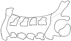
Spinous process suspension method: The exposure is the same as before. First, part of the spinous process is removed to shorten it. Bilateral full-thickness laminotomies are performed near the inner edge of the facet joints. The supraspinous and interspinous ligaments at the lowest level are removed, along with the ligamentum flavum. A bone groove is made on the adjacent spinous process near the lowest level. The lowest spinous process is then sutured with wire or silk thread to the bone groove on the adjacent spinous process to achieve bony fusion, with fat grafts placed on both sides (Figure 1).

Figure 1 Spinous process suspension technique
Myelopathic cervical spondylosis is primarily caused by spinal cord compression symptoms due to cervical herniation of intervertebral disc or osteophytes, commonly occurring in individuals aged 40–60. Numbness and heaviness initially appear in the lower limbs, followed by walking difficulties, potentially leading to spastic paralysis. Neck stiffness and backward extension of the neck can easily cause limb numbness. Tendon reflexes are hyperactive, with positive Hoffmann's and Babinski's signs. Sensory disturbances are often irregular. Superficial reflexes are mostly weakened or absent, while deep sensation remains intact. In severe cases, incontinence of urine may occur. Lateral and anteroposterior X-rays show cervical spine straightening or backward angulation, multiple narrowed intervertebral spaces, and osteophyte formation, particularly at the posterior edges of vertebral bodies. Flexion-extension lateral cervical spine films may reveal cervical instability. CT and MRI can visualize spinal canal stenosis and cervical spinal cord compression or lesions.
Ossification of the posterior longitudinal ligament of the cervical spine progresses slowly, with neck stiffness and limited mobility. Its clinical manifestations closely resemble cervical spondylosis, making diagnosis based solely on symptoms and signs difficult; imaging is essential. Plain X-rays confirm 80% of cases, showing linear or cloud-like ossified shadows along the anterior cervical canal wall. Tomography may further aid diagnosis if needed. CT scans confirm the diagnosis and assess the morphology, distribution, and relationship of ossified tissue to the cervical spinal cord. For diagnosis, MRI images are less definitive than CT scans.
Cervical spinal cord tumors manifest as progressive spinal cord compression, with symptoms worsening from single limbs to all four limbs. Urinary retention and bedridden conditions may occur. Sensory and motor disturbances appear simultaneously. Plain X-rays may reveal enlarged intervertebral foramina, thinned pedicles, widened interpedicular distances, or vertebral/pedicle destruction. For extramedullary intradural tumors, myelography shows a cup-shaped defect. Cerebrospinal fluid protein levels are significantly elevated. CT or MRI aids differential diagnosis.
Syringomyelia predominantly affects young people with a slow progression. Dissociation of pain/temperature sensation and touch sensation is notable, especially marked temperature sensation reduction or loss. Myelography shows no obstruction. MRI confirms the diagnosis, revealing cystic changes and central canal dilation in the cervical spinal cord.
Amyotrophic lateral sclerosis is a motor neuron disease, with symptoms progressing from the upper limbs to the lower limbs, leading to progressive and rigid paralysis. No sensory disturbances or bladder symptoms are present. The spinal canal's sagittal diameter is typically normal, and myelography shows no obstruction.


