| disease | Orbital Blowout Fracture |
| alias | Orbital Floor Fracture |
A blow-out fracture, also known as an orbital floor fracture, was first reported by Lang in 1889, who described a case involving enophthalmos and diplopia caused by an orbital floor fracture. Later, King (1944) reported similar cases, and Smith (1957) formally named it the blow-out fracture.
bubble_chart Etiology
According to Crumley's (1977) statistics on 324 cases of injury causes, car accidents accounted for 65%, boxing 16%, blunt force injuries 11%, falls 6%, and other injuries 2%.
The mechanism of its occurrence is as follows:
(1) The theory of sudden increase in intraorbital pressure: When the anterior part of the eye is struck by a blunt object, the intraorbital tissues are compressed towards the orbital apex, causing a sharp rise in intraocular pressure. This pressure is transmitted to the orbital walls, leading to a fracture at the weak points of the orbital wall. This can cause intraorbital soft tissues such as periorbital fat, the inferior rectus, and the inferior oblique muscles to herniate into the maxillary sinus and become incarcerated.
Cramer et al. (1965) classified the severity of orbital floor fractures into the following five types based on the severity of the trauma:
1. Linear type: No displacement of the fracture fragments.
2. Trapdoor type: The displaced bone fragment often remains connected on the medial side, while the other end protrudes into the maxillary sinus, resembling a trapdoor.
3. Hanging type: The fracture becomes multiple fragments, causing the orbital floor to sag like a hammock.
4. Depressed type: The fracture fragments fall into the maxillary sinus.
5. Complete detachment of the orbital floor.
(2) The theory of orbital wall bending: In 1974, Fujino proposed this theory through experiments with a mechanical model of the orbital region, suggesting that a sudden increase in intraorbital pressure cannot immediately cause an orbital floor fracture. The external force acting on the orbital rim first causes a transient deformation and bending of the entire orbital wall, which then leads to a fracture. Imaging diagnosis and medial orbital wall fractures support this theory. The author believes that this theory is essentially an extension of the theory of sudden increase in intraorbital pressure and can be considered as one and the same.bubble_chart Pathological Changes
The orbital floor slopes downward from the inside to the outside, so the lowest part of the orbital floor is located in a depression about 3 mm anterior and lateral to it. Among the anteroposterior diameters of the orbital walls, this area is the shortest, averaging 47 mm. The majority of the orbital floor is composed of the orbital plate of the maxilla and the orbital surface of the zygomatic bone, each occupying about half of the orbital floor. Additionally, a small portion is formed by the orbital process of the palatine bone.
Between the orbital plate of the maxilla and the orbital surface of the zygomatic bone lies the infraorbital groove, which connects posteriorly with the infraorbital fissure and anteriorly forms the infraorbital canal. Its external opening is located about 4 mm below the infraorbital margin, through which the infraorbital nerve and infraorbital artery pass. Therefore, fractures of the orbital floor often result in numbness of the cheek. The infraorbital groove is closest to the medial side of the infraorbital fissure, where the bone wall is thinnest, at 1-3 mm, making it a common site for fractures.
The inferior rectus muscle is close to the orbital floor, but in the anterior part of the orbital floor, it is separated by the inferior oblique muscle and intraorbital fat. The nerve supplying the inferior rectus muscle enters its upper part at the junction of the middle and posterior third of the muscle. Therefore, in most cases of orbital floor fractures, the nerve of the inferior rectus muscle is not easily injured, but the inferior oblique and inferior rectus muscles are often affected.The lamina papyracea of the ethmoid bone, which forms the medial wall of the orbit, is the thinnest, measuring 0.2-0.4 mm. Thus, fractures of the orbital floor are often accompanied by fractures of the medial orbital wall.
bubble_chart Clinical Manifestations
1. Local symptoms: eyelid swelling, subcutaneous static blood, subconjunctival hemorrhage, subcutaneous lowering qi swelling, and intraorbital emphysema.
2. Diplopia: caused by the entrapment of the inferior rectus muscle in the fracture gap. This symptom appears when looking upward and often occurs after the acute reaction subsides. Downward displacement of the eyeball is also one of the causes of diplopia.
3. Downward displacement of the eyeball: caused by the prolapse of intraorbital soft tissue into the maxillary sinus. A straight line drawn horizontally in front of the eyes can show that the pupil on the injured side is lower than that on the healthy side.
4. Enophthalmos: initially presents as exophthalmos due to intraorbital edema and hemorrhage, and enophthalmos appears several days after the injury when the reaction subsides. It is mainly caused by the enlargement of the orbital cavity and the herniation of intraorbital fat into the maxillary sinus. It is also significantly related to advanced stage lesions such as infection and necrosis of intraorbital fat, retrobulbar adhesions, and scar shortening of the extraocular muscles.
5. Restricted eye movement: often involves restricted movement along the vertical axis of the eye, and the mechanism is still inconclusive. The theory of sudden increase in intraorbital pressure suggests that it is caused by the entrapment of the inferior rectus muscle at the fracture site. Koonreef (1982) based on anatomical studies, believes that eye movement disorders are caused by hemorrhage and swelling of the connective tissue around the extraocular muscles, leading to nerve dysfunction. Hammerschlag (1982) through CT scan analysis found that restricted movement of the inferior rectus muscle is caused by the prolapse of orbital contents pulling and twisting the muscle.6. Numbness in the area of the infraorbital nerve distribution: caused by injury to the infraorbital nerve. The numbness affects the lower eyelid, cheek, ala, and upper lip. This symptom also occurs in fractures of the infraorbital rim and is not unique to blowout fractures. About half of the patients may experience numbness subsiding within a year.
7. Visual impairment: occurs in 20-30% of cases, with the following six causes. Timely examination and diagnosis are necessary to save vision:
(1). Corneal trauma, advanced stage can cause corneal leukoma.
(2). Iris rupture and detachment, iris paralysis, dilated and fixed pupil, advanced stage can cause glaucoma.
(3). Lens dislocation or subluxation, advanced stage can cause internal visual obstruction.
(4). Retinal lesions.
(5). Fracture of the optic canal, optic nerve atrophy.
(6). Penetrating eye injury, which is a contraindication for early orbital floor repair, so careful examination, diagnosis, and timely treatment are necessary.
Classification
Orbital floor fractures often involve fractures in other parts of the face, and there are different classification methods.
I. Converse et al. (1967) classification
1. Orbital floor fracture: ① Simple orbital floor fracture, without injury to the orbital rim. ② Complex orbital floor fracture, with fractures of the orbital rim and face.
2. Polymorphic orbital fracture: ① Linear fracture involving the maxilla and zygoma. ② Comminuted orbital floor fracture with midface fractures. ③ Zygomatic fracture, separation of the frontozygomatic suture, and downward displacement of the zygomatic part of the orbital floor.
II. Crumley et al. (1977) classification
1. Simple orbital floor fracture.
2. Fracture of the orbital rim and floor.
3. Tripod fracture of the zygoma.
4. Central complex medial malleolus fracture (Le Fort II fracture). {|122|}
1. Check the upward movement of the eyeball. If the affected eyeball cannot move upward, the diagnosis can be confirmed.
2. Palpate the orbital rim for any step-like deformity and displacement.
3. Numbness in the distribution area of the infraorbital nerve has reference value.
4. Inferior rectus muscle traction test: Apply surface anesthesia to the conjunctival sac. Use ophthalmic toothed forceps to grasp the inferior rectus tendon from the sclera and rotate the eyeball. If it is incarcerated, the upward movement of the eyeball will be restricted, which can be compared with the healthy side (Figure 1).
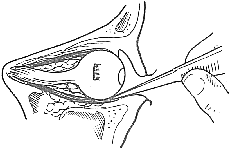
Figure 1 Inferior rectus muscle traction test
Use forceps to grasp the inferior rectus tendon to prove that the upward rotation of the eyeball has recovered, which is negative.
5. X-ray films have important diagnostic value. Taking naso-chin, naso-frontal, and lateral views can reveal the following lesions: ① Abnormal soft tissue shadow at the top of the maxillary sinus. ② Orbital tissue prolapse into the top of the maxillary sinus, showing a hanging drop hammock-like shadow. ③ Sometimes blood and orbital floor bone fragments can be seen protruding into the maxillary sinus. ④ Orbital floor bone defect.
6. Orbital CT scan: Axial and coronal CT scans can clearly show the fracture status and the degree of orbital content prolapse, and can also display other facial fractures, providing a comprehensive evaluation of the patient's injury.
bubble_chart Treatment Measures
Regarding the early implementation of surgical treatment, there was controversy in the past, but a consensus has gradually been reached. If enophthalmos, diplopia, and inferior rectus muscle entrapment are detected, and X-rays show orbital floor destruction, it is advisable to observe for one week until the orbital swelling subsides before performing surgery. The entrapped inferior rectus muscle should be released, and the orbital soft tissues that have prolapsed into the maxillary sinus should be repositioned, along with the reduction of the orbital floor fracture. If observation exceeds three weeks, bony healing will occur at the injury site, making surgery difficult. Conversely, for simple orbital floor fractures without enophthalmos or diplopia, observation can continue for two weeks. If the aforementioned symptoms are absent, conservative treatment can be pursued. In summary, for those requiring surgical treatment, the earlier the surgery, the better the outcome.
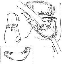
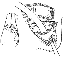
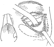
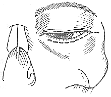
(1) Sub-eyelash incision approach: Under local anesthesia, a transverse incision is made along the natural skin crease below the lower eyelid eyelash. The orbicularis oculi muscle is dissected to the orbital rim. A suture is placed in the middle of the skin incision to retract the skin flap downward, exposing the infraorbital rim. The periosteum is incised transversely, and extensive dissection is performed from the outer aspect of the orbital floor periosteum to explore the orbital floor bone plate. The fracture site is identified, and the entrapped inferior rectus muscle and other orbital tissues are released and pulled back into the orbit. Depending on the size and shape of the orbital floor bone defect, an autologous iliac bone graft is harvested, shaped into a tile-like form, and placed over the bone defect to repair the orbital floor. The surgical procedure must be strictly aseptic (Figure 1).
|
|
|
|
Figure 1: Blowout fracture orbital floor iliac bone graft method
(1) Sub-eyelash incision approach (2) Dissection of the orbital floor, anterior wall of the maxilla, and anterior wall of the zygoma periosteum (3) Dotted line indicates the extent of the subperiosteal pocket (4) Iliac crest bone graft is placed within the pocket, with the outer layer of the bone graft grooved using a drill
(2) Inferior fornix incision approach: Infiltration anesthesia is performed in the inferior fornix, with a small amount of adrenaline added to the local anesthetic to reduce bleeding. An incision is made along the inferior fornix, extending to the medial and lateral canthi. The periosteum of the medial, inferior, and lateral orbital walls is dissected. The orbital floor bone defect is explored, and the entrapped inferior rectus muscle is released and repositioned along with the orbital soft tissues into the orbit. Orbital floor repair is performed based on the specific situation. This approach fully exposes the orbital floor, facilitating the procedure and avoiding facial scarring.
(3) Maxillary sinus approach: The anesthesia and incision are the same as for a radical maxillary sinusectomy. The anterior wall of the maxillary sinus is chiseled open, and blood clots within the sinus are aspirated. The superior aspect of the maxillary sinus is explored, and a suture is placed around the entrapped inferior rectus muscle to assist in dissection. The freed inferior rectus muscle is then pushed upward into the orbit, and the fracture fragments are reduced as much as possible. A medial wall antrostomy is created, and iodoform gauze is packed through it for support and fixation. The packing should remain in place for 10-15 days. This method facilitates the repositioning of orbital tissues and leaves no facial scar, but it is less convenient for orbital floor repair compared to the above methods.
(4) Orbitomaxillary Sinus Combined Approach - Also known as the naso-ocular combined approach, this method was developed by Kirkegaard (1986) based on the principle of complementing strengths and compensating for weaknesses. It combines the aforementioned second and third methods to facilitate the operation and improve therapeutic outcomes.


