| disease | Fracture of the Odontoid Process of the Axis |
Odontoid fractures are not uncommon, accounting for 10-15% of adult cervical vertebral fracture-dislocations. Unfortunately, there are still reports of odontoid fractures being misdiagnosed as fistula disease during initial visits. Any patient with persistent neck pain and stiffness after trauma, with or without symptoms of nerve compression, should undergo repeated X-ray examinations, including CT scans, to avoid missing a possible odontoid fracture.
bubble_chart Pathogenesis
There are several different classification systems for odontoid fractures. Schatzker et al. divided them into high and low types based on whether the fracture line is above or below the accessory ligament. Althoff classified odontoid fractures into four types: A, B, C, and D. Type A fractures pass through the isthmus of the odontoid, while the fracture lines of the other three types are located at lower anatomical positions (Figure 1).
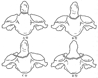
Figure 1: Althoff Classification of Odontoid Fractures
The most widely used clinical classification today is the Anderson and D'Alonzo classification, which divides odontoid fractures into three types: I, II, and III (Figure 2). Type I fractures, also known as apical fractures, are oblique fractures at the attachment sites of the apical ligament and one of the alar ligaments, accounting for about 4%. Type II fractures, or basal fractures, occur at the junction between the odontoid and the body of the axis and are the most common, accounting for about 65%. Type III fractures involve the body of the axis, with a large cancellous bone base below the fracture line, often extending to one or both superior articular surfaces of the axis, accounting for about 31%. Most authors believe this classification is clinically significant, as it helps guide treatment selection and prognosis assessment when combined with factors such as the degree and direction of medial malleolus fractures and the patient's age. However, for Type II odontoid fractures, some authors have proposed subtypes: Hadley et al. introduced Type IIA fractures, defined as basal fractures with a large free bone fragment at the fracture site, representing inherent instability. Pederson and Kostuik proposed Type IIB and IIC fractures. Type IIB fractures correspond to Anderson and D'Alonzo's Type II and Althoff's Type B fractures, while Type IIC fractures are defined as fractures where the fracture line is located above the accessory ligament on at least one or both sides, equivalent to Althoff's Type A fractures (Figure 3).
Additionally, there is a special type of odontoid fracture: epiphyseal separation. The odontoid process of the axis develops a secondary ossification center at its apex around age 2, which fuses with the main part of the odontoid by age 12. The odontoid itself begins to fuse with the body of the axis at age 4, with most fusions completing by age 7. Therefore, before age 7, odontoid fractures typically present as epiphyseal separations.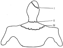
Figure 2: Anderson-D'Alonzo Classification of Odontoid Fractures
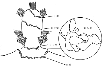
Figure 3: Pederson-Kostuik Classification of Odontoid Fractures
There are also rare reports of vertical odontoid fractures. To date, only two such cases have been reported in English literature: one by Johuson et al. in 1986 and another by Bergenheim et al. in 1991, neither of which fits into the above classifications.
Odontoid fractures are evidently caused by various injury mechanisms. Althoff conducted biomechanical studies on cadaveric cervical spine specimens, applying loads such as hyperflexion, hyperextension, and horizontal shear to the atlantoaxial joint, none of which resulted in an odontoid fracture. Therefore, he concluded that anterior-posterior horizontal forces primarily cause ligamentous damage rather than odontoid fractures. In further experimental studies, the types of loads that caused odontoid fractures, ranked from least to most severe, were: horizontal shear + axial compression, a 45° anterior or posterior oblique impact relative to the sagittal plane, and lateral impact. Thus, he proposed that the combined effect of horizontal shear and axial compression is the primary mechanism for odontoid fractures, while lateral impact is the necessary external force for causing Type A (Type IIC) odontoid fractures. Mouradian et al. also found in experiments that lateral loading can lead to odontoid fractures. Doherty et al., through biomechanical experiments, suggested that lateral or oblique lateral loads result in Type II odontoid fractures, whereas hyperextension forces lead to Type III odontoid fractures. However, clinically, some patients describe injury mechanisms that differ from these findings. Pederson reported a case of a 77-year-old male patient who sustained an anterior-to-posterior force to the frontotemporal region, resulting in a Type IIC odontoid fracture with posterior displacement of up to 20 mm. The injury mechanism in this patient can be hypothesized as a hyperextension force transmitted through the anterior arch of the atlas to the odontoid, causing fracture and displacement. One direct force vector was an anterior-to-posterior vector transmitted through the skull to the anterior arch of the atlas and then to the odontoid, creating a horizontal shear force (Figure 4).
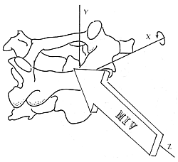
Figure 4 Horizontal shear forces cause the odontoid process to rotate around the X-axis
Odontoid fractures can also occur in flexion-type injuries, resulting in anterior displacement. In this guillotine-like mechanism, an intact transverse ligament is sufficient to transmit enough energy to cause an odontoid fracture and anterior displacement (Figure 5). Under the combined action of multiple forces, the presence of torsional forces makes the odontoid process more prone to fracture. The mechanisms are as follows: (1) During rotation, the alar ligaments are already maximally stretched; (2) During rotation, both ligaments and muscles are under tension, the facet joints are tightly engaged, and injuries in other planes are minimized; (3) The atlantoaxial joint accounts for 50% of cervical rotation, and this region bears the greatest load when subjected to rotational forces. In summary, the mechanism of odontoid fracture is complex, involving flexion, extension, lateral bending, and rotational forces. By analyzing the fracture type, displacement, and the relationship with associated head and facial injuries in a patient, the injury mechanism can often be inferred.
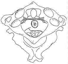
Figure 5 Guillotine mechanism
The transverse ligament transmits energy, causing an odontoid fracture
bubble_chart Clinical Manifestations
Occipital and posterior neck pain are the most common clinical symptoms, often accompanied by radiating pain in the distribution area of the greater occipital nerve. Neck stiffness leads to a forced posture, and the typical sign is the patient supporting their head with their hands to relieve pain, although this scenario is relatively uncommon in clinical practice. Approximately 15–33% of patients exhibit neurological symptoms and signs, with grade I paraplegia and neuralgia being the most frequent. There have been reports of odontoid fractures accompanied by paralysis of the tenth and twelfth cranial nerves. The severity of symptoms depends on the degree and location of spinal cord compression caused by fracture displacement. In severe cases, respiratory arrest may occur, particularly in elderly individuals, often resulting in immediate death.
The clinical manifestations of old odontoid fractures are often insidious, as the history of trauma may sometimes be unclear. Crockard et al. reported a group of 16 patients with old odontoid fractures, among whom 3 had forgotten their history of neck trauma, while the rest were initially misdiagnosed due to physicians underestimating the significance of their trauma. Symptoms included C2 nerve root pain, weakness in both hands, and difficulty walking.bubble_chart Auxiliary Examination
Imaging Examination
For patients with suspected diagnosis, conventional X-ray examination is the first choice, including anteroposterior, open-mouth, and lateral flexion-extension views of the cervical spine. However, due to neck stiffness or even forced posture at the time of consultation, standard and clear X-ray films are sometimes difficult to obtain in one attempt. If the initial X-ray examination does not reveal clear anatomical relationships or definitive signs of fracture, but clinical suspicion remains, two open-mouth views and two occipitocervical lateral views should be considered routine examinations to confirm the diagnosis. However, due to overlapping bony structures in the occipitocervical region, conventional X-ray examinations may occasionally yield negative results when an odontoid fracture is nondisplaced. Therefore, tomograms in sagittal and coronal planes are required under the following circumstances: (1) clinical suspicion of odontoid fracture with negative conventional X-rays; (2) conventional X-rays suggesting suspicious signs of fracture, which is the most common indication; (3) confirmed odontoid fracture but with suspected adjacent concomitant fractures. In Ehara et al.'s imaging study of odontoid fractures, the results of tomograms were classified into three categories: I. Tomograms confirmed the findings of conventional X-rays; II. Tomograms revealed additional findings or provided a more comprehensive presentation of the injury; III. Odontoid fracture was visible only on tomograms. Among their 50 patients with odontoid fractures, 24 were definitively diagnosed on initial conventional X-rays, while the remaining 26 underwent coronal and sagittal tomograms. Among the 19 patients in Group I, two had fractures unconfirmed on coronal tomograms but confirmed on sagittal tomograms. Among the four patients in Group II, two showed fracture lines extending to the superior articular facet of the axis, one showed fracture lines involving the vertebral body of the axis, and one had essentially negative conventional X-rays except for slight deviation of the odontoid on the open-mouth view, with an oblique Type II odontoid fracture visible on coronal tomograms. Another three patients had completely negative conventional X-rays, with fractures visible only on tomograms (Group III). The author has encountered cases in outpatient clinics where initial X-rays suggested suspected odontoid fractures, but two open-mouth views ruled out the diagnosis. Clearly, whether false-positive or false-negative, such results cause harm to patients and should be noted.
X-ray findings of odontoid fractures primarily include bony discontinuity, displacement, and angulation, with displacement being the most reliable sign. Sometimes, lateral angulation of the odontoid on open-mouth views is the only sign. A high-quality lateral view is essential for diagnosing odontoid fractures, as they often involve anteroposterior displacement and angulation, and the direction of displacement has therapeutic implications. However, occasional anatomical variations, such as posterior tilting of the odontoid, should not be misdiagnosed as fractures. Indirect signs, such as prevertebral soft tissue shadows, may only help localize the injury, and sometimes the prevertebral soft tissues appear normal, especially in immediate post-injury examinations. Conversely, head or facial fractures can also cause prevertebral soft tissue swelling. In Ehara's study of 50 patients, 43 (86%) showed fracture signs on an initial lateral view, three showed odontoid fractures on open-mouth views, one was detected on a skull lateral view, and only three had negative conventional X-rays. Tomograms primarily reveal cortical discontinuity and basal shadowing of the odontoid. Sagittal tomograms can demonstrate displacement and angulation of odontoid fractures, providing significant diagnostic value. Harris et al. described a ring-like shadow at the base of the odontoid on sagittal projections, indicating bony discontinuity, as a sign of Type III odontoid fracture.
CT scans can clearly display fractures and displacement, especially when the patient's forced position causes unclear anatomical structures on conventional X-rays. Blacksin and Lee reported 100 cases of severe trauma patients who underwent routine cervical spine lateral radiographs and conventional occipital CT scans as a substitute for open-mouth views. The results showed that CT detected 7 cases with 8 fractures, 3 located at the foramen magnum of the occipital bone and 5 at the C12 level. In contrast, conventional cervical spine X-rays only revealed widened prevertebral soft tissue shadows in 2 cases and failed to identify any fracture signs, highlighting the importance and superiority of CT examinations. MRI can clearly show spinal cord compression caused by fracture displacement, the extent of spinal cord injury, and the condition of adjacent soft tissue injuries. Pederson reported a case of odontoid fracture diagnosed by MRI 16 weeks post-injury due to persistent neck pain and progressive neurological compression symptoms. The MRI revealed a complete fracture of the atlantoaxial complex with posterior dislocation and severe spinal cord compression. Upon reviewing the X-rays, it was found that the cervical spine lateral radiograph taken on the day of injury showed an odontoid fracture, with the fracture line passing through the isthmus (classified as a Type IIC fracture) and the fractured end displaced posteriorly by approximately 20mm. Subsequent CT scans confirmed the 20mm posterior displacement of the odontoid fracture, demonstrating no change in fracture displacement 16 weeks post-injury.
A detailed and accurate history of injury and physical examination often enables physicians to consider the possibility of this injury. Motorcycle accidents are a common cause of odontoid fractures in younger populations, while in the elderly, the most frequent cause of this injury is a simple fall. An odontoid fracture with posterior dislocation is a more severe injury than one with anterior dislocation, with a higher likelihood of neurological symptoms, and is more common in the elderly population.
X-ray examination is the primary basis and means for diagnosing odontoid fractures. When the diagnosis is in doubt, repeated imaging, tomography, or CT scans should be performed. MRI can provide information on spinal cord injury. In cross-section, the odontoid process and the spinal cord each occupy one-third of the sagittal diameter of the spinal canal, with the remaining one-third serving as a buffer space (Figure 1). The distance between the posterior edge of the anterior arch of the atlas and the odontoid process (AO interval) in adults is 2mm to 3mm, slightly larger in children at 3mm to 4mm. Exceeding this range should raise suspicion of an odontoid fracture and/or ligamentous disruption. Asymmetry of the odontoid process on an open-mouth view should also raise suspicion of injury in this area. A clear open-mouth view can reveal the odontoid fracture and its type. A lateral view can show the fracture type, anterior or posterior displacement, and whether there is dislocation of the atlantoaxial vertebrae. Additionally, attention should be paid to any associated deformities or fractures in the occipitocervical region.
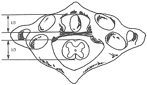
Figure 1: The odontoid process and spinal cord each occupy one-third of the sagittal diameter of the spinal canal.
The diagnosis should be differentiated from transverse ligament rupture, transverse ligament avulsion, and atlantoaxial posterior dislocation. In transverse ligament rupture, the AO interval exceeds 5mm, and the odontoid process remains intact. In transverse ligament avulsion, irregular bone fragments may be seen between the lateral masses of the atlas on an open-mouth view, and a CT scan can confirm the diagnosis by revealing small defects in the lateral mass surface of the atlas and free bone fragments. Atlantoaxial posterior dislocation on a lateral X-ray shows an inverted position of the anterior arch and the odontoid process, with occasional small fracture fragments anterior or at the tip of the odontoid. Additionally, attention should be paid to any associated occipitocervical deformities, such as occipitalization of the atlas or basilar invagination.
The diagnosis of an odontoid fracture should include the following five points: (1) the type of odontoid fracture; (2) the presence and direction of displacement; (3) the presence of neurological injury; (4) the presence of associated injuries to adjacent bones and soft tissues; (5) the presence of concomitant injuries in other parts of the body.
bubble_chart Treatment Measures
The nonunion rate of untreated or improperly treated odontoid fractures ranges from 41.7% to 72%, accompanied by potential atlantoaxial instability. Once displacement occurs, it may lead to acute or chronic injury of the brainstem, spinal cord, or nerve roots, resulting in severe limb paralysis, respiratory dysfunction, or even death. Therefore, patients with odontoid fractures should receive active treatment, with comprehensive consideration of fracture type, displacement, age, and other factors to adopt appropriate therapeutic measures.
Non-surgical treatment includes direct plaster fixation, traction reduction + plaster fixation, and Halo brace fixation. For stable fractures without displacement, direct plaster fixation can be chosen, followed by radiographic review after 8–12 weeks. After clinical healing, a cervical collar should be used for protection for an additional 2–3 months. For odontoid fractures with displacement, traction reduction + plaster fixation is employed. The traction weight is generally 1.5–2 kg and should not be excessive to avoid over-traction, which may lead to nonunion. The direction of traction and neck position should be adjusted based on the displacement of the fracture. Repeated radiographic reviews (bedside films including anteroposterior and lateral views) should be conducted within 2–3 days to assess reduction status and adjust traction positioning. Once satisfactory reduction is achieved, maintain neutral positioning with continued traction for 3–4 weeks, followed by supine head-neck-thorax plaster fixation while maintaining traction. Remove the plaster after 3–4 months and take X-rays to evaluate fracture healing. After clinical healing, follow the same protocol as above. Premature plaster fixation may lead to nonunion. Halo ring fixation of the head, connected to a thoracic plaster via support rods, provides considerable stability. Foreign literature reports that it can restrict 86% of cervical motion, yielding favorable therapeutic outcomes. However, its installation is complex, and complications such as pin-site infections and pressure sores are not uncommon due to perforation and fixation. Additionally, Anderson et al. conducted a prospective study of 42 cervical injury patients immobilized with Halo braces, taking supine and upright lateral X-rays within 5 days of Halo brace fixation post-injury. In uninjured segments, positional changes resulted in an average angulation of 3.9°, with the greatest motion occurring between the occipital bone and atlas (8°). In injured segments, the average sagittal angulation was 7°, with an average horizontal displacement of 1.7 mm. The range of motion in injured segments was unrelated to the injury level or fracture type. Among 45 injured planes, 35 exhibited angulation greater than 3° or displacement exceeding 1 mm, accounting for 77%. Therefore, the authors recommend that supine and upright lateral X-rays should be taken when using Halo braces to treat unstable cervical injuries. If excessive motion is observed, alternative treatments should be considered. Halo brace fixation also lasts 3–4 months, followed by cervical collar protection for 2–3 months after radiographic confirmation of fracture healing.
Surgical treatment includes anterior screw compression internal fixation between fracture ends, posterior fusion, and decompression at sites of spinal cord compression.
1. Anterior screw fixation for fracture end compression: The currently popular anterior screw fixation techniques are generally similar, involving drilling from the anteroinferior aspect of the axis vertebral body to the apex of the odontoid process. A standard cortical lag screw uses a 2.5mm long drill bit, while a cannulated screw uses a 1.2mm Kirschner wire, reaching the posterior half of the cortical bone at the apex of the odontoid process. The hole is then tapped, and a screw of appropriate length is inserted (Figure 1). The entire procedure must be performed under simultaneous biplanar fluoroscopic guidance to immediately confirm the direction and depth of the Kirschner wire and screw, as well as the position of the fracture ends. During drilling and tapping, it is absolutely necessary to retract and protect the soft tissues to prevent injury to critical structures. The screw should reach the cortical bone at the posterior half of the odontoid apex but must not penetrate the cortex into the foramen magnum of the occipital bone. Debates arise regarding the choice and number of screws. Rilger et al. suggest that using two double-threaded screws can provide compression and rotational stability at the fracture site. McBride conducted biomechanical studies on cadaver specimens, comparing the strength of two 3.5mm cannulated AO screws versus one 4.5mm cannulated Herbert screw for odontoid fracture fixation. The average torsional strength was 1196 N·m/deg for the 4.5mm Herbert screw group and 434 N·m/deg for the 3.5mm AO screw group, showing a significant difference. The shear strength was 106.9 and 86.1 kN/m2 (K·N/M2), respectively, with no significant difference. They concluded that one 4.5mm cannulated Herbert screw provides superior stability for type II odontoid fractures compared to two 3.5mm cannulated AO screws, while also reducing surgical time and risk by requiring only one screw. Graziaro et al. compared one versus two 3.5mm screws for odontoid fracture fixation in eight cadaver specimens and found similar results: no significant difference in bending or torsional strength between one and two screws. Sasso et al. reported comparable findings: although two screws provided higher resistance to extension, the load required to cause fixation failure was not significantly different. Additionally, they found that screw fixation provided only 50% of the stability of an intact odontoid. Doherty similarly concluded that single-screw fixation achieves half the stability of an unfractured odontoid. Knoringer noted that double-threaded screws, with their shafts almost entirely embedded in bone, simplify the procedure, reduce surgical risk, and cause less postoperative soft tissue irritation. Nucci et al. measured 92 normal adult odontoids and determined that placing two 3.5mm odontoid screws requires at least a 9.0mm inner diameter, concluding that 95% of subjects met this size requirement. Chang et al. achieved 100% union rates by using a single 4.5mm double-threaded screw for non-displaced or anatomically reducible type II odontoid fractures and a single 3.0mm double-threaded screw for partially reducible fractures, with satisfactory outcomes.
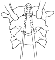
Figure 1 Odontoid fracture anterior screw internal fixation surgery
First, drill a hole with a 2.5mm drill bit, then tap the thread with a 3.5mm tap
Postoperative management: Observe in the ICU for 24 hours post-surgery, closely monitoring respiratory status. Wear a rigid cervical collar for protection for 6 weeks, removing it only during rest and bathing. Follow-up X-rays should be taken at 6 weeks, 12 weeks, and 24 a cycle of day and night post-surgery.
Contraindications for anterior screw internal fixation: (1) Odontoid fracture with unilateral or bilateral atlantoaxial joint fracture; (2) Odontoid fracture with unstable Jefferson fracture; (3) Unstable type III odontoid fracture, unsuitable for Halo brace or gypsum fixation; (4) Atypical type II odontoid fracture: comminuted fracture or fracture line oblique and nearly parallel to the intended screw insertion direction; (5) C1~2irreversible fracture displacement, such as old fracture. (6) Odontoid fracture with transverse ligament rupture; (7) Unstable type II fracture or shallow type III fracture, accompanied by significant kyphosis deformity limiting cervical extension; (8) Unstable type II fracture or type III fracture in elderly patients with degenerative spinal canal stenosis; (9) Pathological odontoid fracture (Figure 2).
From a biomechanical perspective of the spine, anterior screw compression internal fixation between fracture ends is superior to posterior fusion, adhering to AO/ASIF principles. Additionally, it preserves at least partial upper cervical rotation function, which is a significant advantage. However, improper technique or use in contraindicated cases can lead to numerous complications. This procedure requires specialized instruments and dual "C"-arm fluoroscopy, which are expensive and currently difficult to widely adopt domestically.
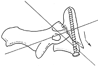
Figure 2 Oblique fracture line is a contraindication for anterior screw fixation
2. Posterior fusion: Includes upper cervical posterior fusion and occipitocervical fusion.
Upper cervical posterior fusion includes wire fixation (Gallie and Brooks-Jenkins techniques) and transarticular screw fixation. The former has been introduced in the previous chapter; here, we discuss transarticular screw fixation.
Surgical method: The patient is placed in a prone position, and lateral fluoroscopy confirms the reduction of the odontoid fracture. The neck is flexed to facilitate screw placement, and fluoroscopy is repeated before disinfection to ensure no re-displacement.
A posterior midline incision is made from the occipital protuberance to C4, exposing the posterior arch of the atlas, and the spinous processes, laminae, and articular processes of C2~3. For residual irreducible anterior dislocation, gentle traction on the axis spinous process and atlas posterior arch can achieve reduction. Note: Rebound clamping can be fatal, so clamping must be secure. For residual irreducible posterior dislocation, stress reduction can be attempted. Remember, reduction should not be forced or performed violently.
The sharp knife meticulously dissects the axis lamina and articular process. The upper part of the lamina and isthmus is stripped with a sharp nerve dissector, exposing upward to the posterior joint capsule of the atlantoaxial joint, while avoiding exposure of the lateral vertebral stirred pulse to prevent injury. The screw insertion point is made 2 mm lateral to the inner surface of the articular process and 3 mm above the edge of the inferior articular process. Under lateral fluoroscopic guidance, a 2.5 mm long drill bit is advanced in a completely sagittal direction, entering the lateral mass from the medial part of the isthmus and penetrating the cortex of the atlas lateral mass anteriorly. The length is measured, followed by tapping with a 3.5 mm cortical bone tap, and then the screw is inserted. The entire procedure is performed under lateral fluoroscopic guidance to avoid drilling in a horizontal direction, which could prevent entry into the atlas lateral mass and risk injuring the vertebral stirred pulse.
After the insertion of bilateral screws, perform C1–2 posterior fusion. The use of bone grafting and posterior wire fixation methods can enhance the stability of fixation and the fusion rate. In cases where the posterior arch of the atlas is fractured or decompression is performed, atlantoaxial joint fusion should be conducted. Use a sharp nerve dissector to retract the soft tissue containing the greater occipital nerve, exposing the atlantoaxial joint. Insert a Kirschner wire into the lateral mass of the atlas to serve as both traction and a marker. Incise the joint capsule to expose the atlantoaxial joint, then use a small sharp osteotome to remove the posterior half of the articular cartilage, fill it with cancellous bone, and secure it with compression screws.
Postoperative management: Same as that for anterior screw fixation.
Transarticular screw fixation is biomechanically superior to wire fixation and is suitable for acute or chronic atlantoaxial instability, particularly when accompanied by a fracture of the posterior arch of the atlas or when C1 posterior decompression is required. This method can avoid the need for occipitocervical fusion (see the previous chapter), although it presents certain technical challenges.
Selection of Treatment Methods
The choice of treatment should be based on a comprehensive consideration of factors such as the type of fracture, presence of displacement, reduction status, and age.
Epiphyseal separation occurs exclusively in children under 7 years old and typically does not present neurological symptoms. The preferred treatment is conservative management. Posterior cervical fusion is considered only when traction fails to achieve or maintain reduction.
Type I odontoid fractures are generally stable fractures. Since the fracture site is distant from the transverse ligament, even nonunion under insufficient immobilization does not result in instability, making conservative treatment appropriate. However, some authors argue that Type I odontoid fractures include: (1) rupture of at least one alar ligament's occipital bone attachment; (2) partial rupture of the apical ligament, potentially serving as a radiographic sign of atlanto-occipital instability. These fractures are considered unstable and pose a life-threatening risk, possibly requiring respiratory and hemodynamic support, necessitating close monitoring during management. If longitudinal separation is present, immediate immobilization with a Halo brace is required. For cases with anterior or posterior displacement, skull traction is employed to achieve reduction and decompression. Most patients require occipitocervical fusion to achieve stability.
Type II odontoid fractures are the most common and present significant treatment challenges, with considerable debate. Conservative treatment has a high nonunion rate, reported as 36% by Anderson and D'Alonzo. The currently recommended approach involves skull traction for reduction, followed by posterior fusion and decompression as needed. Indications for posterior fusion include: (1) cervical spinal cord injury; (2) persistent cervical symptoms; (3) severe fracture displacement (≥5 mm) or angulation deformity (≥10°); (4) atlanto-dens interval greater than 5 mm; (5) old fractures or nonunion.
For Type II odontoid fractures with posterior displacement, the Gallie technique may lead to redisplacement, making the Brooks-Jenkins technique preferable. In the Gallie method, wires are looped around the posterior arch of the atlas and the spinous process of the axis, then tied behind a posterior bone graft, generating a backward force on the atlas that can cause postoperative redisplacement. In contrast, the Brooks-Jenkins technique uses two wedge-shaped bone grafts placed between the posterior arch of the atlas and the lamina of the axis on each side, secured with compression wires. This method avoids backward force and helps maintain reduction. Additionally, patients with posterior displacement are more likely to have associated posterior atlantoaxial fractures, which should be carefully assessed preoperatively. The optimal choice in such cases is posterior transarticular screw fixation.
For patients with nonunion or old fracture of type II odontoid fracture, the conventional method is posterior fusion. Some authors have selectively achieved success using anterior screw internal fixation. For patients with cervical spinal cord compression, it is necessary to determine whether the compression originates from the anterior or posterior. For primarily anterior compression, indirect posterior decompression cannot relieve the pressure, and an anterior transoral approach is required to remove the odontoid for decompression, combined with various posterior fusion techniques.
Type III odontoid fracture A nondisplaced type III fracture is stable and can be treated with plaster and cervical collar immobilization. For displaced type III fractures, traction reduction followed by plaster immobilization may be performed. Foreign literature often recommends halo vest treatment, aiming to correct angular deformities with the halo vest and immobilize until fracture healing. Significant anterior displacement or angular deformity of the odontoid can lead to spinal canal stenosis and spinal cord compression, but residual displacement generally does not require treatment. For some special unstable fractures, surgical treatment should be considered, including posterior fusion and, occasionally, anterior screw internal fixation.
Age Age over 60 is considered an indicator of poor healing for odontoid fractures, especially type II fractures, and surgical treatment should be considered. Bednar et al. conducted a prospective study on 11 odontoid fracture patients with an average age of 74 and found that aggressive treatment (early surgery and early postoperative mobilization) significantly reduced mortality in elderly odontoid fracture patients.
Multiple injuries and associated injuries For patients with multiple injuries, comprehensive diagnosis is necessary to prioritize and manage conditions in sequence. For patients with congenital anomalies, such as platybasia or occipitalization of the atlas, the location of spinal cord compression and the nature of the injury should be fully evaluated based on imaging studies, considering the trauma mechanism and spinal cord function, to achieve decompression and stabilization.
Treatment protocol
Given the current domestic situation, anterior screw fixation is still difficult to popularize, despite being a treatment method more aligned with spinal biomechanics. For a patient with an odontoid fracture, the first step is to establish a clear diagnosis, with definitive answers to the five diagnostic criteria. For patients with associated injuries to adjacent bones and ligaments, early surgical treatment may be considered. For those without associated injuries to adjacent tissues, conservative treatment should be prioritized, aiming for early traction reduction, followed by maintained traction and immobilization to promote healing. For patients with difficult reduction or failure to maintain reduction, as well as nonunion in old fractures, early atlantoaxial fixation and fusion may be considered. For patients with atlantoaxial instability, often accompanied by spinal cord and nerve compression symptoms, simple atlantoaxial fixation is insufficient. Decompression by removing the posterior arch of the atlas is necessary, and in some cases, the posterior rim of the occipital bone's foramen magnum should also be excised for decompression, followed by occipitocervical fusion.
Fuju et al. proposed a treatment algorithm for odontoid fractures, combining literature and the authors' experience. The diagram is as follows: (Figure 3)
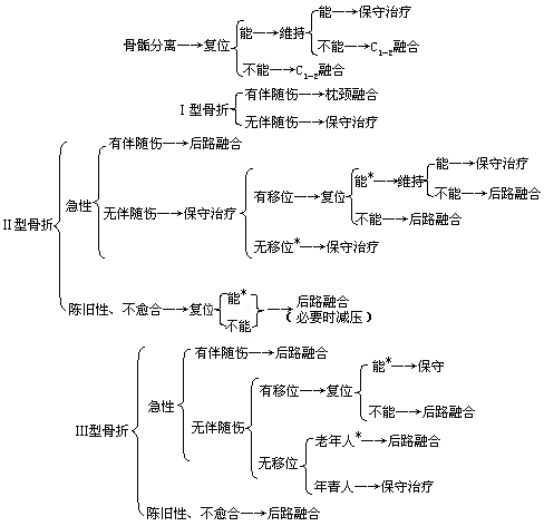
Figure 3 Treatment algorithm for odontoid fracture
Note: 1. Associated injuries refer to injuries of adjacent bones and ligaments that cause fracture instability; 2. Cases marked with * may also undergo anterior screw fixation.


