| disease | Deviated Nasal Septum |
| alias | Structural Rhinitis |
Deviation of the nasal septum refers to an intranasal deformity where the nasal septum deviates from the midline and causes clinical symptoms. In fact, most people have some degree of nasal septum deviation, but whether it causes nasal symptoms often depends on the following factors: ① the degree and form of deviation, such as a prominent localized protrusion that happens to be in the nasal valve area; ② the extent of turbinate transformation; ③ whether there are deformities in the bone at the lateral edge of the pear-shaped aperture or the cartilage in the nasal valve area.
bubble_chart Etiology
1. Trauma Trauma is the most common cause of nasal septum deviation. When a nasal bone fracture occurs due to trauma, it is often accompanied by dislocation and deformation of the septal cartilage, or even cartilage fracture. If reduction is insufficient during recovery, it may result in residual septum deviation. During childbirth, a narrow birth canal or improper use of forceps can also lead to septal cartilage deviation or dislocation. Gray found that among 2095 cases of normal childbirth, nasal septum deviation accounted for 4%. However, if the final stage of childbirth lasted more than 15 minutes, the injury rate of nasal septum deviation could reach 13%, and it was believed that one-third of nasal septum dislocations occurred during the rotation of the fetal head within the pelvis. Sooknumlun (1986) reported that among 201 newborns, 31 cases had nasal septum deviation, accounting for 15.4%.
2. Enlarged adenoids in children affect nasal ventilation, leading to compensatory mouth breathing. Over time, this can cause developmental deformities of the maxillofacial bones and a high-arched hard palate, resulting in an elevated nasal floor and gradually leading to a deviated septum.
3. The uneven growth rates of different parts of the nasal septum can lead to deformities, which are more likely to occur at the junctions between these parts.bubble_chart Clinical Manifestations
Depending on the degree and location of the deviation, the following symptoms may occur:
1. Stuffy nose — This is the most common symptom of nasal septum deviation, often presenting as persistent stuffy nose. If the deviation is unilateral, it results in one-sided stuffy nose, whereas an "S"-shaped deviation may cause bilateral stuffy nose.
2. Epistaxis — The protruding area of the deviation has thin and fragile mucosa, which, when irritated by inhaled airflow over time, can lead to inflammatory reactions and nosebleeds. Such bleeding is usually minor and typically occurs in the anterior part of the septum.
3. Reflexive headache — If the deviated portion is in contact with or presses against the middle or inferior turbinate, it often causes ipsilateral headache and may also contribute to nasal neuralgia. The headache may lessen or disappear after using nasal vasoconstrictors or surface anesthesia of the nasal mucosa.
4. Symptoms involving adjacent structures — If the deviated part of the septum corresponds to the middle meatus or middle turbinate, it may compress and displace the middle turbinate outward or cause excessive pneumatization and mucosal hypertrophy of the middle turbinate, obstructing sinus drainage in the middle meatus. Over time, this can lead to sinusitis and associated symptoms.
Recently, Low (1993) conducted tympanometric measurements on 40 patients with nasal septum deviation-induced stuffy nose and found significantly reduced middle ear pressure on the affected side. After surgical correction of the deviation, middle ear pressure improved. Clinically, patients often present with unilateral ear fullness or hearing loss.
Examination — Anterior rhinoscopy can generally reveal the type and degree of deviation, but posterior septal deviations often require careful examination to detect.
Septal deviations can be classified by direction as a "C"-shape (unilateral) or an "S"-shape (bilateral), and by morphology as a ridge (semi-circular protrusion) or a spur (sharp protrusion). Trauma-induced dislocation of the septal cartilage may sometimes protrude into the nasal vestibule.
High deviations of the septum often tightly contact the middle turbinate, narrowing the middle meatus. In cases of significant septal deviation, the size difference between the two nasal cavities is noticeable. When one nasal cavity is significantly narrowed, compensatory hypertrophy of the contralateral turbinate is often observed.
The diagnosis can usually be made through nasal endoscopy. However, it must be differentiated from a nasal septum nodule. The latter occurs in the high posterior part of the septum near the middle turbinate and is a protrusion formed by localized thickening of the septal mucosa, which feels soft when probed. The formation of septal nodules is related to chronic irritation from purulent nasal discharge. Another rare condition is a syphilitic gumma of the nasal septum, which is also relatively firm in texture, but the mucosa in that area is markedly congested.
bubble_chart Treatment Measures
Surgical correction is the only treatment method. However, if there are concurrent nasal polyps or turbinate hypertrophy, nasal polyp and turbinate surgery should be performed first. If nasal ventilation improves and nasal symptoms disappear, the deviated septum may not require treatment. Surgery should be performed if any of the following conditions are present: ① Persistent long-term stuffy nose caused by septal deviation. ② High septal deviation affecting sinus drainage. ③ Recurrent epistaxis due to septal deviation. ④ Reflex headache caused by septal deviation. ⑤ Vasomotor rhinitis (structural rhinitis) with significant septal deviation.
(1) Submucous resection of the nasal septum This is the most common method for treating septal deviation. The patient is placed in a semi-sitting position. Cotton pads soaked in 1% dicaine (with a small amount of 0.1% adrenaline) are placed in both olfactory clefts and the common nasal meatus for surface anesthesia of the nasal mucosa. Then, infiltration anesthesia is performed at the incision site of the anterior septum using 1% procaine or 0.5% lidocaine with 0.1% adrenaline, which facilitates the separation of the mucoperichondrium and mucoperiosteum. Injecting a small amount of saline under the mucoperichondrium of the nasal septum to create a bulge can aid in the separation, especially at sharp cartilaginous spurs.
1. Incision An incision is made at the junction of the left nasal vestibule skin and the septal mucosa, extending from the top of the anterior septum to the base of the septum, with appropriate extension toward the nasal floor for easier surgical manipulation. Care must be taken not to cut through the cartilage to avoid injury to the contralateral mucoperichondrium. The soft tissue over the cartilage is incised to expose the porcelain-white cartilage.
2. A septal elevator is inserted under the mucoperichondrium at the incision site, and the mucoperichondrium and mucoperiosteum are separated closely along the cartilage surface. The separation proceeds from top to bottom parallel to the nasal bridge, primarily relying on the side edge of the elevator. The separation range extends anteriorly and superiorly to the upper edge of the septal cartilage and inferiorly to the base of the septum. The mucoperiosteum at the nasal floor must also be partially separated, extending upward and backward beyond the deviated portion by at least 1 cm.
3. When reaching a deviated protrusion, careful separation should first be performed around the protrusion to reduce mucosal tension before finally separating the mucoperiosteum at the protrusion.
4. Approximately 2 mm posterior to the original incision, the septal cartilage is incised with a septal knife, but the contralateral mucoperichondrium must not be cut through. The blade should be angled obliquely to the cartilage plane.
5. The elevator is inserted through the cartilage incision to the contralateral mucoperichondrium and is used to separate the contralateral mucoperichondrium and mucoperiosteum closely along the cartilage surface, with the same range of separation as the incision side. Extra caution is required when separating the concave area of the deviated cartilage, as the mucoperichondrium may adhere tightly to the depressed cartilage, and care must be taken to prevent soft tissue rupture. To facilitate separation at the deepest concavity, a small amount of saline can be injected under the mucoperichondrium before separating the soft tissue to create a bulge, aiding in the separation.
6. After separating the tissues on both sides of the septal cartilage and bone, a nasal septum speculum is inserted through the incision to spread the soft tissues on both sides, fully exposing the cartilaginous and bony surfaces of the septum. A septal rotary knife is used to first push the perpendicular plate of the ethmoid bone backward parallel to the nasal bridge from the upper end of the cartilage incision, then downward to the vomer, and finally forward along the anterior-superior edge of the vomer and the nasal crest of the maxilla, removing most of the septal cartilage. The removed cartilage is temporarily preserved in case of soft tissue rupture on either side, where it can be placed flat between the ruptured soft tissues to prevent future septal perforation. When using the rotary knife, care must be taken not to remove too much of the anterior-superior edge of the septum, leaving at least 6 mm of cartilage to prevent future nasal bridge collapse.
7. Use a nasal septum rongeur to remove the deviated portion of the bony nasal septum. For the bony ridge at the base, a nasal septum osteotome can be used for removal. Regarding the perpendicular plate of the ethmoid bone, excessive removal should be avoided, and forceful twisting to fracture the bone should not be performed to prevent injury to the cribriform plate and subsequent complications.
8. Remove blood, blood clots, and bone debris generated during the procedure thoroughly, then extract the nasal septum spreader. Ensure the bilateral soft tissues adhere properly and inspect both nasal cavities to confirm the correction of the deviated septum. If any residual deviation remains, reinsert the spreader to address it.
9. After the bilateral soft tissues adhere, suture the incision with 2–3 stitches or leave it unsutured. Pack both nasal cavities with sterile Vaseline gauze. During packing, first lay the gauze over the incision or any ruptured soft tissue for protection, then fill the remaining space with additional gauze.
10. Remove the nasal gauze after 24 hours. To prevent sneezing post-removal, instruct the patient to gently press the lower edges of both nasal bones with their fingernails. Postoperatively, instill 1% Ephedrine saline drops into the nose daily for one week, and use Ephedrine-soaked cotton pads to shrink and clean the nasal cavity every other day. If the mucoperichondrium or mucoperiosteum is ruptured during surgery, take extra care during postoperative cleaning to promptly remove excess secretions and prevent infection. If sutures are present, remove them after five days.
(II) Septoplasty The advantage of septoplasty is its ability to correct deformities, straighten the nasal septum, and preserve the septal cartilage framework, with a very low risk of postoperative septal perforation. This procedure is suitable for deviations in the cartilaginous portion of the nasal septum, with contraindications similar to submucous resection of the nasal septum.
1. Anesthesia and patient positioning are the same as for submucous resection of the nasal septum.
2. Incision On the concave side of the deviated septum, retract the columella toward the opposite side. Make a vertical incision from top to bottom along the free edge of the septal cartilage in the nasal vestibular skin, extending from the nasal dorsum to the anterior nasal spine, fully exposing the anterior edge of the septum and the nasal spine.
3. From the incision, dissect backward to separate the soft tissue and underlying mucoperiosteum on the concave side of the septum, fully exposing the septal framework on the incised side.
4. Detach the septal cartilage from the nasal dorsum and the upper lateral cartilage. Remove a narrow strip (3–4 mm wide) of septal cartilage from the posterior-superior and to be decocted later inferior junctions with the bone, leaving the entire septal cartilage attached only to the contralateral undissected flap tissue.
5. If the bony framework of the septum is also deviated, insert a septal elevator through the posterior edge of the detached septal cartilage to separate the mucoperiosteum on the contralateral bony septum. Then, use a septal punch forceps to remove the deviated portions of the perpendicular plate of the ethmoid bone, vomer, etc.
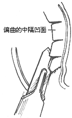
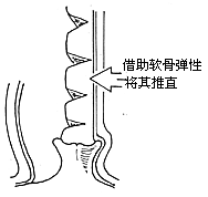
6. At the most prominent concave area of the septal cartilage, make several deep incisions on the concave side of the cartilage surface without penetrating the contralateral mucoperichondrium. The incisions should extend nearly to the entire edge of the cartilage depression. Alternatively, excise 1 mm-wide strips of cartilage between the incisions. The elasticity of the treated cartilage will allow it to straighten, and it can then be repositioned to the midline (Figure 1).


Figure 1 Septoplasty
(1) Making incisions on the concave side of the deviated septum (2) Using cartilage elasticity to straighten it
7. Inspect both nasal cavities for any remaining deviations. If residual deviations are found, reopen the incision for further correction.
8. Suture the incision and pack both nasal cavities with Vaseline gauze under equal pressure. Remove the gauze after 24 hours. Other postoperative measures are the same as for submucous resection of the nasal septum.
1. Perforation of the Nasal Septum ① Most often occurs during the surgical separation of soft tissues due to rough handling, causing bilateral ruptures at symmetrical locations. ② When the nasal mucosa has infectious inflammation, even a unilateral tear can lead to perforation. ③ If the soft tissues on both sides of the nasal septum are inadequately separated, removing cartilage or bone can easily tear the soft tissues. In cases of bilateral tearing, the extracted cartilage fragment can be placed between the two ruptures, and a "U"-shaped plastic sheet can be placed over the surface of the bilateral ruptures for protection. Then, both sides are evenly packed with Vaseline gauze, and the plastic sheet is carefully removed afterward.
2. Hematoma of the Nasal Septum Caused by bleeding between the nasal septal cartilage and mucosa. If semicircular bulges are found on both sides of the nasal septum after removing the nasal packing post-surgery, with a soft texture, and the patient complains of a stuffy nose that does not improve even with Ephedrine saline drops, a small hematoma may resolve on its own, while a larger one requires intervention.
3. Abscess of the Nasal Septum Results from bacterial infection of a nasal septal hematoma. Improper timing of surgery, such as during an intranasal infection or when there is significant purulent discharge, can lead to infection at the incision site.
4. Collapse of the Nasal Bridge Often caused by excessive removal of nasal bridge cartilage during surgery. If postoperative nasal septal abscess is not properly managed, leading to cartilage softening and necrosis, this deformity may also occur.
5. Intracranial Complications Rare. A nasal septal abscess or damage to the cribriform plate can increase the risk of intracranial infection. {|104|}


