| disease | Pulmonary Hamartoma (Surgery) |
Pulmonary hamartoma ranks first in incidence among benign lung tumors.
bubble_chart Etiology
The origin and cause of pulmonary hamartoma are not yet fully understood. A widely accepted hypothesis suggests that a hamartoma arises from a fragment of bronchial tissue that becomes inverted and detached during embryonic development, subsequently being enveloped by normal lung tissue. This displaced tissue grows slowly or may remain dormant for a period before eventually developing into a tumor. The fact that most hamartomas occur after the age of 40 supports this hypothesis.
bubble_chart Pathological Changes
Hamartomatous tumor diseases are characterized by abnormal combinations and arrangements of normal tissues. This histological anomaly may involve disorganization in the quantity, structure, or maturation level of organ tissues. The main tissue components of hamartomas include cartilage, fat, smooth muscle, glands, epithelial cells, and sometimes bone tissue or calcification. There have been no reports of malignant transformation in hamartomas.
Hamartomas are generally solid, dense, spherical, or oval in shape, but can also be lobulated or nodular, with most measuring less than 3cm in diameter.
bubble_chart Clinical Manifestations
Hamartomas mostly develop in individuals over the age of 40, with males being more affected than females.
The vast majority of hamartomas (over 80%) grow in the peripheral areas of the lungs, closely attached beneath the visceral pleura, and sometimes protrude from the lung surface. As a result, they generally present no clinical symptoms and show no positive signs during physical examinations. Only when a hamartoma grows to a certain size, large enough to irritate or compress a bronchus, causing bronchial stenosis or obstruction, do clinical symptoms such as cough, chest pain, fever, shortness of breath, bloody sputum, or even hemoptysis appear. At this stage, corresponding clinical signs, such as wheezing or tubular breathing sounds, may also be observed.
bubble_chart DiagnosisThe diagnosis of hamartoma mainly relies on X-ray examination, and most cases are incidentally discovered during routine X-ray checks. On X-rays, it appears as a uniformly dense shadow, which can also be uneven, and may exhibit calcification. The calcification pattern resembles popcorn, with relatively lower density at the periphery, possibly due to fatty tissue. The popcorn sign is a characteristic feature of pulmonary hamartoma, though it is uncommon and not exclusive to pulmonary hamartoma (Figure 1).
Pulmonary hamartomas are generally solitary, with multiple occurrences being extremely rare and not yet reported domestically. The vast majority of solitary hamartomas are of the intraparenchymal lung type, while the endobronchial type is very uncommon. They occur more frequently in the right lung than the left, and more often in the lower lobes than the upper lobes, with some cases occurring in the right middle lobe and the lingular segment of the left upper lobe.
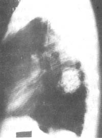
Figure 1 Pulmonary hamartoma
bubble_chart Treatment Measures
Pulmonary hamartomas detected during health check-ups can be extremely difficult to differentiate from malignant lung tumors due to the lack of dynamic observation. Rapidly enlarging pulmonary hamartomas in the short term are also challenging to diagnose definitively. Therefore, when clinical and X-ray findings cannot rule out malignancy, surgery should be performed as early as possible. Even for benign hamartomas, early surgical intervention can prevent complications such as pneumonia, atelectasis, and bronchiectasis caused by tumor enlargement, which may worsen or complicate the condition.
The surgery is performed under general anesthesia. Upon opening the chest, the tumor is visible on the lung surface, with a firm texture and an irregular surface. The tumor can also be felt sliding within the lung tissue. The tumor can be completely excised by slightly separating the lung tissue after incision. Except for endobronchial hamartomas or cases where malignancy cannot be ruled out, local resection or segmental resection is generally performed.


