| disease | Malignant Tumors of the Ear |
| alias | Adenoid Cystic Carcinoma of External Auditory Canal, Basal Cell Carcinoma, Melanoma, Squamous Cell Carcinoma |
smart_toy
bubble_chart Overview Malignant tumors occurring in the external ear are relatively rare. According to foreign statistics, malignant tumors of the auricle and its adjacent structures account for approximately 6% of all cutaneous malignancies. Friedmann (1974) reported 295 cases of ear malignancies, of which 58 cases (19%) involved the auricle, 158 cases (54%) the external auditory canal, and 79 cases (72%) the middle ear. Regardless of whether they occur in the auricle or the external auditory canal, squamous cell carcinoma is the most common type of malignant tumor in the external ear. The next most common types are basal cell carcinoma in the auricle and adenoid cystic carcinoma in the external auditory canal. Other malignant tumors, such as rhabdomyosarcoma and malignant melanoma, are extremely rare.
bubble_chart Etiology
The disease cause of squamous cell carcinoma:
is not very clear. Squamous cell carcinoma of the auricle may be related to ultraviolet radiation, such as prolonged exposure to strong sunlight. Squamous cell carcinoma of the external auditory canal may be associated with inflammatory stimulation from chronic external otitis or chronic otitis media.
bubble_chart Clinical Manifestations
1. Clinical manifestations of squamous cell carcinoma:
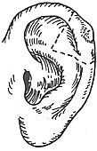
In the early stage, squamous cell carcinoma of the auricle presents as scaly patches or papules with itching, which may bleed easily upon scratching. It gradually develops into hard nodules, followed by surface erosion, ulceration, or the formation of cauliflower-like masses. In the initial stage [first stage], there is no pain, but in the advanced stage, when the cartilage membrane is involved, the pain becomes more pronounced. Squamous cell carcinoma of the external auditory canal is often misdiagnosed as chronic external otitis or sebaceous cyst in the early stages. Patients frequently present with bloody ear discharge (fistula disease). Examination may reveal localized skin erosion in the external auditory canal with granulation-like tissue growth. A biopsy can usually confirm the diagnosis. Auricular squamous cell carcinoma progresses slowly, and metastasis occurs relatively late. The most common sites of metastasis are the parotid lymph nodes, followed by the jugulodigastric lymph nodes and the upper posterior cervical lymph nodes. Squamous cell carcinoma of the external auditory canal often grows invasively, quickly invading underlying bone tissue and potentially affecting the facial nerve.
2. Clinical manifestations of basal cell carcinoma:
Initially, it often appears as a small gray nodule or slightly raised hard patch on the skin, causing no discomfort. Occasionally, itching may occur, and scratching may lead to bleeding (water from river). The hard nodule gradually enlarges, with the center ulcerating to form an ulcer and the edges becoming raised, resembling a crater. The tumor grows invasively but generally progresses slowly. Metastasis of basal cell carcinoma is rare. When metastasis occurs, it primarily spreads via local lymph nodes, though distant metastasis may also occur, commonly to the lungs and bones.
3. Clinical manifestations of adenoid cystic carcinoma of the external auditory canal:
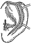
Adenoid cystic carcinoma of the ceruminous glands in the external auditory canal grows very slowly, with a history that may span several years before diagnosis. In the early stages, intermittent ear pain is common, which may progress to persistent severe pain in the advanced stage, radiating to the temporal region and around the ear. Tumor obstruction of the external auditory canal can cause tinnitus and conductive hearing loss. In cases with a prolonged course, secondary infections and ear fistula disease may occur, such as concurrent external otitis or otitis media.
Local examination typically reveals a mass in the cartilaginous portion of the external auditory canal, often located on the anterior-inferior wall. The mass is broad-based, firm, and may be tender. The overlying skin is usually intact (except in cases of infection), and tenderness may be present. The tumor may also present as an annular hard nodule, narrowing the external auditory canal. If the tumor breaks through the skin, it may appear as red granulation tissue, with bloody or purulent discharge visible in the external auditory canal.
4. Clinical manifestations of melanoma:
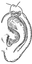
Melanoma is more common in middle-aged and elderly individuals. The tumor often occurs on the helix, concha, or may be found in the external auditory canal and retroauricular region. Early lesions are flat, smooth, and exhibit gray-black pigmentation. In the advanced stage, a mass forms, accompanied by ulceration and necrosis. It is important to note that if a benign pigmented nevus of the ear shows accelerated growth, a burning sensation, pain, or surface erosion and bleeding, there should be high suspicion of malignant transformation into melanoma.
bubble_chart Diagnosis
Diagnosis of adenoid cystic carcinoma of the external auditory canal:
This tumor is often clinically misdiagnosed as external auditory canalitis, otitis media, or exostosis of the external auditory canal. Therefore, special attention should be paid to middle-aged or elderly patients with long-term painful masses in the external auditory canal, particularly those with significant ear pain in the early stages of the disease but without local acute inflammatory manifestations. The final diagnosis must rely on pathological examination.
bubble_chart Treatment Measures
I. Treatment of Squamous Cell Carcinoma:
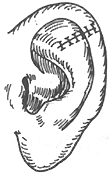
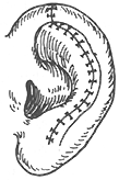
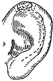
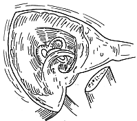
The primary approach is surgical excision. For squamous cell carcinoma of the auricle, different surgical methods can be employed depending on the tumor's location and size. Smaller tumors located on the helix can be excised and sutured using wedge-shaped or star-shaped incisions during the initial stage [first stage]. For larger tumors, a posterior auricular advancement flap can be used in two stages to repair the auricle defect post-excision. If the tumor involves most of the auricle, total auriculectomy and split-thickness skin grafting are required (Figures 1 and 2). For localized squamous cell carcinoma of the external auditory canal with a small tumor size, en bloc excision of the external auditory canal can be performed. The excision should include the skin of the external auditory canal, the surrounding bony walls, the tympanic membrane, and the malleus. For extensive tumors invading adjacent tissues with cervical lymph node metastasis, modified temporal bone resection and neck dissection are necessary. In some cases, the parotid gland and temporomandibular joint may also need to be excised (Figure 3). Radiotherapy is less effective for squamous cell carcinoma of the external ear and should not be used alone but can be combined with surgical treatment.
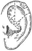
Figure 1 Surgery for Small Auricle Carcinoma
(1)(2) Wedge excision (3)(4) Star-shaped excision (5)(6)(7) Wide excision with flap reconstruction
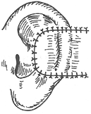
Figure 2 Extensive auricle carcinoma resection
(1)(2)(3)(4) Partial auricle resection (5)(6) Total auricle resection with skin grafting
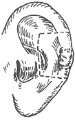
Figure 3 External auditory canal carcinoma resection
(1) Partial resection of the outer or inner segment of the external auditory canal carcinoma; (2) Bony segment resection; (3) Surgical field after extensive resection
II. Treatment of basal cell carcinoma:
Surgical resection is the primary approach, which may be combined with radiotherapy. If the tumor involves a wide range, such as invading cartilage, the external auditory canal, or the middle ear, extensive temporal bone resection is required.
III. Treatment of adenoid cystic carcinoma of the external auditory canal:
This tumor is pathologically often low-grade malignant but lacks a capsule, exhibiting infiltrative growth. Local recurrence is highly likely postoperatively, and the prognosis is poor. Early extensive resection is recommended. For cases with a short disease duration and localized tumor extent, total external auditory canal resection may be performed. The resection scope should include cartilage, the bony external auditory canal, the tympanic annulus, the tympanic membrane, the malleus, mastoid air cells, and the zygomatic arch root. If necessary, parotidectomy may also be performed. For cases with extensive involvement, subtotal or total temporal bone resection, including condylar process resection of the mandible and upper cervical lymph node dissection, should be performed. Radiotherapy may benefit some patients, but generally, this tumor is poorly sensitive to radiotherapy.
IV. Treatment of melanoma:
Early surgical resection is the primary approach. For small superficial melanomas on the helix, wedge resection may be performed. For infiltrative growth with larger tumors, auricle resection, parotidectomy, and neck lymph node dissection should be considered based on the extent of involvement. Melanoma is insensitive to radiation.