| disease | Hypersplenism |
| alias | Hypersplenism, Hypersplenism |
Hypersplenism, commonly referred to as splenomegaly, is a syndrome characterized by an enlarged spleen, a reduction in one or more types of blood cells, and corresponding hyperplasia of bone marrow hematopoietic cells. Blood counts typically improve and symptoms alleviate after splenectomy.
bubble_chart Etiology
Hypersplenism can be divided into two major categories: primary and secondary:
(1) Primary hypersplenism: Includes so-called primary splenic hyperplasia, non-tropical idiopathic splenomegaly, primary splenic granulocytopenia, primary splenic pancytopenia, splenic anemia, or splenic thrombocytopenia. Since the disease cause is unknown, it is difficult to determine whether this group of diseases represents different outcomes caused by the same disease cause or unrelated independent diseases.
(2) Secondary hypersplenism: Occurs in cases with the following clearly identified disease causes: ① Acute infections with splenomegaly, such as viral hepatitis or infectious mononucleosis; ② Chronic infections, such as subcutaneous nodules, brucellosis, malaria, etc.; ③ Congestive splenomegaly due to portal hypertension, including intrahepatic obstruction (e.g., portal cirrhosis, postnecrotic cirrhosis, biliary cirrhosis, hemosiderosis, sarcoidosis, etc.) and extrahepatic obstruction (e.g., external compression or thrombosis of the portal or splenic veins); ④ Inflammatory granulomas such as systemic lupus erythematosus, rheumatoid arthritis, Felty syndrome, and sarcoidosis; ⑤ Malignant tumors such as lymphoma, leukemia, and metastatic carcinoma; ⑥ Chronic hemolytic diseases such as hereditary spherocytosis, autoimmune hemolytic anemia, and thalassemia; ⑦ Lipid storage diseases such as Gaucher disease and Niemann-Pick disease; ⑧ Myeloproliferative disorders such as polycythemia vera, chronic granulocytic leukemia, and myelofibrosis; ⑨ Other conditions include splenic hemangioma and cavernous hemangioma.
(3) Occult hypersplenism: Whether primary or secondary hypersplenism, due to good compensatory bone marrow hyperplasia, peripheral blood counts may not show cytopenia. However, once factors such as infection or drugs suppress hematopoietic function, it can lead to single or pancytopenia.The structure of the spleen
The spleen is composed of connective tissue framework, lymphoid tissue, blood vessels, lymphatic vessels, and cells of the hematopoietic and mononuclear-macrophage systems. The spleen is divided into white pulp and red pulp, with an intermediate transitional zone called the marginal zone, which is the area where the red pulp receives the stirred pulse blood (Figure 1). The white pulp consists of dense lymphoid tissue and is the main distribution area of T lymphocytes, resembling the structure of lymphoid follicles but with a rich blood circulation. The small stirred pulse branches from the trabecular stirred pulse are surrounded by abundant lymphoid nodules, known as the central stirred pulse of the lymphoid sheath. The lymphoid sheath contains densely packed lymphocytes and plasma cells. The central stirred pulse and its branch small stirred pulses are perpendicular, so most of the branch small stirred pulses contain plasma with few blood cells, which is beneficial for the spleen's immune function. Antigens in the blood pass through the branch small stirred pulses and capillaries to directly contact the lymphocytes and plasma cells in the lymphoid sheath, stimulating the production of more immunologically active cells. Due to antigen stimulation, germinal centers may appear in the white pulp, containing proliferating B cells that can produce corresponding antibodies.

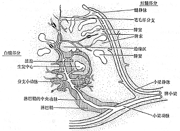
Figure 1 Structure and blood circulation of the spleen
The red pulp contains many irregular fissures called splenic sinuses. The sinus walls are composed of endothelial cells, basement membrane, and outer membrane reticular cells, with small pores in the basement membrane that allow blood cells to pass through easily. Between the sinuses are the splenic cords. The walls of the splenic cords consist of a layer of reticular cells and endothelial macrophages. The terminal ends of the central stirred pulse enter the red pulp and divide into many non-communicating fine penicillar branches. Most penicillar branches open directly into the splenic cords (open circulation), while a few connect with the sinuses (closed circulation). The blood in the penicillar branches is more concentrated, with increased viscosity, causing the blood cells in the splenic cords to move slowly and remain in the cords for an extended period. Combined with the spleen's unique blood circulation structure, blood cells find it difficult to quickly pass through the basement membrane pores to reach the splenic sinuses (Figure 2).
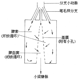
Figure 2 Relationship between open and closed circulation in the spleen, indicating the direction of blood flow
Functions of the spleen
(1) Retention of blood cells: Blood cells are easily retained in the splenic cords of the red pulp. Aging red cells move slowly through the tortuous splenic cords, especially under conditions of low glucose and an acidic environment, gradually becoming spherical with increased osmotic fragility. They cannot pass through the basement membrane pores into the splenic sinuses and are eventually phagocytized by macrophages in the center of the splenic cords. Normal platelets have increased adhesiveness in the splenic cords and are easily trapped by the reticular fibers. After injecting 51

(2) Immune function: The spleen is the largest lymphoid organ in the body. During infections, allergic reactions, and autoimmune diseases, some of the antibodies produced by the body originate from the spleen. The spleen is the primary site for IgM production. Antigens in the blood enter the red pulp, are phagocytized by macrophages, and reach the germinal centers, leading to antibody production, expansion of the germinal centers, and nuclear division. Experimental animals and children who undergo splenectomy are prone to infections, mostly meningitis, acute myocarditis, or acute endocarditis, with high mortality rates. This demonstrates the spleen's crucial role in immune defense against infections.
(3) Blood Storage The spleen's membrane contains smooth muscle fibers that extend into the parenchyma through trabeculae. Therefore, after exercise, acute blood loss, or adrenaline injection, the spleen can undergo rhythmic contraction and relaxation. In normal individuals, the spleen is small in size and has a limited blood storage capacity, estimated to be only about 20 ml. However, when the spleen is significantly enlarged, its blood storage capacity increases, even reaching up to 20% of the total blood volume, playing a regulatory role in systemic blood flow.
(4) Hematopoiesis and Regulatory Functions of the Spleen During the embryonic period, the spleen can generate various blood cells, but after birth, it only produces monocytes and lymphocytes. Experiments have shown that pluripotent stem cells (CFU-S) still appear in the splenic circulation. The spleen seems to have a certain influence on the distribution of CFU-S in the blood. The splenic blood pool is the primary location of CFU-S in the blood. Some researchers have even found that erythroid progenitor cells (BFU-E and CFU-E) can migrate from the bone marrow to the spleen. Therefore, under pathological conditions, extramedullary hematopoiesis can occur, and the spleen can once again produce red blood cells, white blood cells, and platelets, such as in cases of myelofibrosis or metastatic marrow cancer. After splenectomy, the white blood cell and platelet counts in peripheral blood can rise rapidly within hours, peaking at 2–3 days and one week, respectively. Blood smears show a significant increase in flat cells and target cells, and sometimes immature red blood cells, sideroblasts, and red blood cells containing Howell-Jolly bodies can be observed. These phenomena suggest that the normal spleen also plays a role in controlling the maturation of blood cells and their release from the bone marrow into the bloodstream.
Pathogenesis:
The mechanisms by which hypersplenism causes cytopenia are summarized as follows:
(1) Excessive Sequestration In healthy individuals, the spleen does not store red or white blood cells, but about one-third of platelets and some lymphocytes are sequestered in the spleen. When the spleen is pathologically enlarged, not only are more platelets (50–90%) and lymphocytes sequestered, but over 30% of red blood cells may also be retained in the spleen, leading to reduced platelet and red blood cell counts in peripheral blood.
(2) Excessive Filtration and Phagocytosis In hypersplenism, the mononuclear-macrophage system in the spleen becomes hyperactive, and abnormal red blood cells (such as spherocytes or those damaged by receptors, oxidants, other chemical toxins, or physical injury) in the splenic cords significantly increase. These cells are cleared by macrophages, resulting in a marked reduction of red blood cells in peripheral blood. Some red blood cells develop Heinz bodies on their membranes or contain Howell-Jolly bodies, or even Plasmodium trophozoites in their cytoplasm. When these cells attempt to pass from the splenic cords into the sinuses, they often become trapped and are eventually culled by sinus-lining macrophages, with their membranes sustaining damage. After repeated damage, the red blood cells become spherocytes and ultimately cannot pass through the basement membrane pores, leading to their phagocytosis.
Additionally, some scholars propose that in hypersplenism, the spleen produces excessive humoral factors that inhibit the release and maturation of hematopoietic cells in the bone marrow. Others suggest that hypersplenism is an autoimmune disorder, but these theories lack strong evidence and require further research for confirmation.
The diagnosis of hypersplenism relies on the following indicators:
(1) Splenomegaly In almost most cases, the spleen is enlarged. For those whose spleen is not palpable below the ribs, further examinations should be conducted to confirm whether it is enlarged. The use of 99mtechnetium, 198gold, or 113mindium colloid injection followed by spleen area scanning helps estimate the size and shape of the spleen. Computed tomography can also measure spleen size and intrasplenic lesions. However, the degree of splenomegaly does not necessarily correlate with the severity of hypersplenism.
(2) Cytopenia Red blood cells, white blood cells, or platelets may decrease individually or simultaneously. In general early-stage cases, only white blood cells or platelets are reduced, while advanced-stage cases may present with pancytopenia.
(3) Bone marrow shows hematopoietic cell hyperplasia Some cases may also exhibit maturation disorders, or the massive destruction of peripheral blood cells and excessive release of mature cells may create a similar appearance of maturation disorder.
(4) Changes after splenectomy Splenectomy can bring blood cell counts close to or back to normal, unless bone marrow hematopoietic function has already been impaired.
(5) Radionuclide scanning After injecting 51Cr-labeled platelets or red blood cells into the body, surface scanning reveals that the amount of 51Cr in the spleen area is 2–3 times greater than in the liver, suggesting excessive destruction of platelets or red blood cells in the spleen.
When considering the diagnosis of hypersplenism, the first three indicators are particularly important.
bubble_chart Treatment Measures
Splenectomy and X-ray radiation therapy cannot eliminate the primary disease causing hypersplenism, so the primary disease should generally be treated first. If this is ineffective, then active treatment of the primary disease should follow after splenectomy. The indications for splenectomy include the following points:
1. Significant splenomegaly causing obvious compressive symptoms.
2. Severe anemia, especially in cases of hemolytic anemia.
3. Considerable thrombocytopenia with bleeding symptoms. If the platelet count is normal or shows grade I reduction, thrombocytosis may occur after splenectomy, and even thrombosis may develop. Therefore, splenectomy is not advisable for patients with normal or grade I reduced platelet counts.
4. Agranulocytosis with a history of recurrent infections.
Patients undergoing splenectomy should receive thorough preoperative preparation. For example, those with severe anemia should receive blood transfusions, those with thrombocytopenia and bleeding should be treated with corticosteroids, and those with agranulocytosis should take active measures to prevent infections.


