| disease | Acute Cervical Disc Herniation |
Acute cervical disc herniation refers to cases with varying degrees of neck trauma history, imaging evidence confirming disc rupture or protrusion, without cervical vertebral fractures or dislocations, and presenting corresponding clinical symptoms. Before the 1980s, due to limitations in diagnostic technology, understanding of this condition was insufficient, and diagnosis was relatively difficult. Since the advent of magnetic resonance imaging, the detection rate of this condition has progressively increased, and both fundamental and clinical research have continuously advanced.
bubble_chart Etiology
Acute cervical disc herniation is caused by neck trauma. The primary mechanism of injury is acceleration force leading to rapid head movement, resulting in neck sprain, commonly seen in traffic accidents or sports activities. It can be caused by frontal, rear, or lateral impacts, with the most severe disc injuries occurring from extension-acceleration injuries due to rear-end collisions. Generally, acute cervical disc herniation occurs when a certain degree of degenerative changes in the disc is subjected to external force, but it can also occur in discs without obvious pre-existing degeneration.
bubble_chart Pathological Changes
The intervertebral disc is the tissue in the human body that undergoes the earliest and most age-related degenerative changes. With aging, the nucleus pulposus loses some of its water content and its original elasticity. A degenerated cervical intervertebral disc can lead to herniation of intervertebral disc with even minor trauma. Hyperextension injury of the cervical spine can cause the proximal vertebral body to shift backward, while flexion injury can result in bilateral facet joint dislocation, increasing tension in the posterior part of the intervertebral disc and leading to rupture of the annulus fibrosus and posterior longitudinal ligament, with subsequent herniation of the nucleus pulposus.
Taylor, through autopsy studies, pointed out that the most characteristic pathological sign of traumatic cervical intervertebral disc injury is rupture of the cartilaginous endplate, which differs from degenerative changes such as uncovertebral joint fissures and central disc fissures. The cartilaginous endplate fissures in cervical intervertebral discs often appear as linear cracks, close to and parallel with the vertebral endplate, frequently involving the surrounding annulus fibrosus near the vertebral edge, presenting as a "rim lesion." The fissures in the cartilaginous endplate and the lamellar structure of the annulus fibrosus are continuous, often containing hemorrhage, and the nucleus pulposus can herniate through these fissures.Cervical intervertebral disc injuries most commonly occur in the upper three cervical discs. Acute traumatic cervical herniation of intervertebral disc is most frequently seen at the C3–4 level, primarily due to: (1) During cervical hyperextension injury, the shear force is greater, and the C3–4 level is closer to the point of impact compared to the lower cervical vertebrae; (2) The facet joint surfaces at C3–4 are more horizontal, making them more prone to transient anterior-posterior displacement during injury, resembling a flexible joint.
Due to the stabilizing effect of the denticulate ligaments, the cervical spinal cord is relatively fixed. When external forces cause rupture of the intervertebral disc annulus fibrosus and posterior longitudinal ligament, herniation of the nucleus pulposus can easily compress the cervical spinal cord. After compression, the cervical spinal cord becomes thinner and softer, and may form cavities in the early stages. Although the area of spinal cord injury is not large, many patients may exhibit varying degrees of paralysis.
The cervical nerve roots enter the intervertebral foramen horizontally at the level of the intervertebral disc. The posterolateral annulus fibrosus and posterior longitudinal ligament of the cervical spine are relatively weak, making it easier for the nucleus pulposus to herniate in this region. Even small herniations can compress the nerve roots.bubble_chart Clinical Manifestations
The disease has an acute onset, with most cases having a clear history of head and neck trauma. Some cases may be triggered by minor injuries or even stretching. Clinical manifestations vary significantly depending on the location and degree of compression. Based on the location of the herniation of intervertebral disc and the compressed tissues, the disease can be classified into three types: lateral, central, and paramedian (Figure 1).
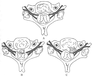
Figure 1 Classification of acute cervical disc herniation
A: Lateral type; B: Central type; C: Paramedian type
1. Lateral cervical disc herniation
The protrusion occurs lateral to the posterior longitudinal ligament and medial to the uncovertebral joint, compressing the cervical nerve roots passing through this area, resulting in radicular compression symptoms.
Symptoms (1) Neck pain, stiffness, and limited movement, resembling "stiff neck"; (2) Severe pain may occur during neck hyperextension, radiating to the scapula or occiput; (3) Pain or numbness in one upper limb, rarely occurring bilaterally simultaneously.
Signs (1) The neck is held in a stiff position; (2) Tenderness and percussion pain at the paravertebral area of the affected segment, with possible tenderness between the lower cervical spinous processes and the medial scapula; (3) Positive cervical nerve root tension test and Spurling test; (4) Sensory, motor, and reflex changes in the affected nerve root distribution area. There may be muscle atrophy and weakness in the innervated muscles.
2. Central cervical disc herniationThe protrusion is located in the central spinal canal, directly anterior to the spinal cord, compressing the bilateral anterior aspects of the spinal cord and causing bilateral spinal cord compression symptoms.
Symptoms (1) Varying degrees of limb weakness, often more severe in the lower limbs than the upper limbs, manifesting as unsteady gait; (2) In severe cases, incomplete or complete paralysis of the limbs may occur; (3) Dysfunction of bowel and bladder, presenting as urinary retention and difficulty defecating.
Signs (1) Varying degrees of limb muscle weakness; (2) Sensory abnormalities, involving both superficial and deep sensations, with the level of sensory disturbance depending on the segment of the herniation of intervertebral disc; (3) Increased muscle tone in the limbs; (4) Hyperreflexia, possible positive patellar and ankle clonus, and positive pathological signs such as Hoffmann and Oppenheim signs.
3. Paramedian cervical disc herniation
The protrusion is located slightly to one side, between the cervical nerve root and the spinal cord, compressing the unilateral nerve root and spinal cord. In addition to the symptoms and signs of the lateral type, there are varying degrees of unilateral spinal cord compression symptoms, manifesting as atypical Brown-Sequard syndrome. This type often presents with severe radicular pain that masks the spinal cord compression symptoms, but once spinal cord compression becomes apparent, the condition is usually severe.
Based on clinical manifestations and imaging findings, the diagnosis is usually not difficult. The diagnostic criteria are as follows:
Medical History
History of head or neck trauma, even a minor neck sprain. The onset is acute, with no symptoms before the onset, followed by signs of cervical spinal cord or nerve root compression.
Imaging Studies
Cervical X-ray: The following may be observed: (1) Reduction or loss of the cervical physiological curvature; (2) In younger patients or those with acute traumatic herniation, the intervertebral space may show no significant abnormalities, but in older individuals, the affected intervertebral space may exhibit varying degrees of degenerative changes; (3) In cases of acute hyperextension injury leading to herniation of the intervertebral disc, the prevertebral soft tissue shadow may appear widened; (4) Dynamic cervical X-rays may sometimes reveal instability in the affected segment.
CT Scan: Although CT scans can provide some diagnostic assistance, conventional CT scans alone are often insufficient for a definitive diagnosis. CTM (myelography combined with CT scanning) can more clearly visualize spinal cord and nerve root compression caused by the intervertebral disc. In recent years, some scholars have advocated the use of this method for diagnosing cervical disc herniation, considering its diagnostic value for lateral cervical disc herniation to be significantly greater than that of MRI.
Magnetic Resonance Imaging (MRI): MRI can directly display the location, type, and extent of cervical intervertebral disc herniation, as well as the degree of spinal cord and nerve root damage, providing reliable evidence for the diagnosis, treatment selection, and prognosis of cervical disc herniation. The diagnostic accuracy of MRI for cervical disc herniation far exceeds that of CT and CTM. For central and paracentral cervical disc herniation, MRI can clearly show: (1) Central type: The intervertebral disc protrudes en masse from the affected intervertebral space, compressing the central anterior portion of the cervical spinal cord. The compressed spinal cord may appear curved, flattened, or indented, with posterior displacement and abnormal signal intensity, predominantly signal enhancement. Sometimes, intramedullary cavitation may be observed; (2) Paracentral type: The intervertebral disc protrudes en masse or in fragments posterolaterally, compressing the lateral aspect of the cervical spinal cord and one nerve root. The anterolateral cervical spinal cord is deformed by compression, displacing posteriorly or toward the unaffected side, with local signal enhancement. The nerve root may be displaced posterolaterally or its image may disappear. Lateral cervical intervertebral disc herniation often requires combined CTM for diagnosis.
Electromyography (EMG)
Used to confirm nerve root damage and has some significance in localizing the affected nerve root. Normal EMG findings indicate preserved nerve root function and a favorable prognosis.
bubble_chart Treatment Measures
Treatment Principles
Non-surgical therapy is the primary approach. If symptoms of spinal cord compression occur, surgical treatment should be performed as early as possible.
Non-surgical therapy
Cervical traction: For herniation of intervertebral disc without degeneration, traction can restore the height of the intervertebral disc, and some protrusions may be reduced. Traction method: Perform in a sitting or lying position using a Glisson band for traction, with a weight of 2.0–3.0 kg. Continuous traction is generally considered more effective than intermittent traction, with a treatment course of 2 weeks. Traction is suitable for lateral cervical disc herniation but should be used cautiously for central cervical disc herniation, as it may worsen the condition.
Cervical collar immobilization: Its main functions are to limit neck movement, enhance neck support, and reduce intradiscal pressure. A simple cervical collar can generally be used for protection. For severe cases with obvious cervical instability, a gypsum collar may be used for fixation. Immobilization is beneficial for recovery after traction-induced symptom relief.
Tuina (massage therapy): Although there are many reports of successful treatment, tuina, especially forceful tuina, may aggravate herniation of intervertebral disc and cause injury to the spinal cord or nerve roots. In severe cases, paralysis may occur instantly during tuina, so caution is advised when using this method.
Physical therapy: This has some effect for mild cases with only nerve root irritation symptoms, with wax therapy and vinegar iontophoresis showing relatively good results.
Medication: Symptomatic treatment, such as sedatives and analgesics, can be used for severe pain.
Surgical therapy
For confirmed cervical disc herniation with severe nerve root or spinal cord compression symptoms, surgical treatment should be performed.
Anterior cervical decompression: Suitable for patients with central or paramedian herniation of intervertebral disc. The use of a trephine for decompression and removal of the injured intervertebral disc, combined with interbody bone grafting and fusion, yields good results. For cases with pre-existing degeneration, osteophytes should also be removed to avoid residual compressive factors.
Posterior cervical decompression: Suitable for lateral cervical disc herniation, multi-segment involvement, or cases accompanied by spinal canal stenosis or ossification of the posterior longitudinal ligament. For simple herniation of intervertebral disc, hemilaminectomy and partial facetectomy can be performed to remove the disc tissue compressing the nerve root through the decompression opening. For cases with spinal canal stenosis or ossification of the posterior longitudinal ligament, total laminectomy may be used.
Cervical microdiscectomy: There are two approaches, posterior and anterior. The choice of approach remains highly controversial in treating soft cervical disc herniation. Aldrich achieved good results using a posterolateral approach to treat lateral nucleus pulposus herniation with single nerve root involvement. The extent of facetectomy during surgery depends on the relationship between the nerve root and the herniated disc. The advantages of this method are: (1) Simple operation; (2) Small incision and minimal trauma; (3) Few complications and low risk. However, this procedure is only suitable for simple cervical disc herniation and is not recommended for cases combined with cervical spinal canal stenosis or ossification of the posterior longitudinal ligament, as the limited decompression scope results in poor surgical outcomes.
Cervical disc nucleolysis: Proposed and first studied by Bonafe and Lazorthes of France. Suitable for cervical disc herniation requiring surgery, especially in young patients who show no improvement after weeks of non-surgical therapy. Although many scholars report that its efficacy is comparable to surgical treatment, several factors limit its widespread use: (1) The anterior cervical puncture approach is used, but the anterior neck has dense anatomical structures such as vascular-nerve bundles and tracheoesophageal bundles, increasing the difficulty and risk of puncture; (2) The use of chymopapain carries a potential risk of spinal cord injury.
1. Cervical spondylosis
There is no clear history of trauma, or symptoms of cervical spondylosis existed before the trauma. The onset is slow, and the symptoms and signs may be similar to those of cervical disc herniation. Imaging shows that osteophytes and intervertebral discs together constitute the compressive lesions, often with the former being the main factor.
2. Intraspinal tumors of the cervical spine
There is no history of trauma, and the onset is generally slow. Imaging can provide important differential evidence. Intramedullary tumors are relatively easy to distinguish, while extramedullary tumors have clear boundaries with the intervertebral discs.
3. Scapulohumeral periarthritis and thoracic outlet syndrome
These mainly need to be differentiated from lateral cervical disc herniation. Scapulohumeral periarthritis only involves shoulder pain and limited movement, without neurological abnormalities. The clinical manifestations of thoracic outlet syndrome closely resemble those of lateral cervical disc herniation, but cervical MRI shows no herniation of the intervertebral disc or nerve root compression. Chest X-rays may reveal narrowing of the thoracic inlet or cervical ribs.


