| disease | Acute Appendicitis |
Acute appendicitis is a common surgical condition, ranking first among various acute abdominal diseases. It was first named by Fitz in 1886, and in 1889, McBurney proposed the concept of surgical treatment for this disease. Over the past century, due to advancements in surgical techniques, anesthesia, antimicrobial therapy, and nursing care, the vast majority of patients have been cured, with the mortality rate dropping to around 0.1%. Common clinical manifestations include migratory right lower abdominal pain and tenderness and rebound pain at McBurney's point. However, the condition of acute appendicitis is highly variable, so each specific case should be taken seriously, with a thorough medical history and careful examination to ensure accurate diagnosis, early surgery, prevention of complications, and improvement of the cure rate.
bubble_chart Etiology
1. Appendiceal lumen obstruction The anatomical characteristics of the appendix, such as a narrow lumen, small opening, abundant lymphoid tissue within the wall, and a short mesentery causing the appendix to curve into an arc shape, are all factors that make the lumen prone to obstruction. Additionally, food residues, fecaliths, foreign bodies, ascarids, tumors, etc., often cause lumen obstruction. After lumen obstruction, the appendix mucosa secretes mucus, forming an abdominal mass, and the intraluminal pressure rises, leading to impaired blood flow, which exacerbates appendicitis.
2. Impact of gastrointestinal diseases Some gastrointestinal diseases, such as acute enteritis, inflammatory bowel disease, schistosomiasis, etc., can directly spread to the appendix or cause spasms of the appendix wall muscles, leading to impaired blood flow and resulting in inflammation.
3. Bacterial invasion After obstruction and inflammation occur in the appendix, the mucosa ulcerates, the epithelium is damaged, and bacteria within the lumen cannot be expelled, taking the opportunity to proliferate and invade the wall, exacerbating the infection. The pathogenic bacteria are mostly various Gram-negative bacilli and anaerobic bacteria from the intestinal tract.Although acute appendicitis often manifests as a suppurative infection caused by varying degrees of bacterial invasion of the appendiceal wall, its mechanism of disease is a relatively complex process, which can be summarized as related to the following factors.
1. Obstruction of the appendiceal lumen: The appendiceal lumen is narrow and elongated, with a closed distal end. Obstruction of the lumen is the basis for inducing acute appendicitis. After the appendiceal lumen is obstructed, a large amount of mucus accumulates in the lumen, causing the intraluminal pressure to gradually rise. Excessive pressure can compress the mucous membrane, leading to necrosis and ulceration, creating conditions for bacterial invasion. If the intraluminal pressure continues to increase, the appendiceal wall is also compressed, first obstructing venous return, leading to venous thrombosis, edema, and ischemia of the appendiceal wall, allowing bacteria in the lumen to penetrate into the abdominal cavity. In severe cases, the blood supply is also obstructed, causing partial or even complete necrosis of the appendix.
The site of appendiceal lumen obstruction is mostly at the root of the appendix, but it can also occur in the middle and distal segments. The causes of obstruction include:
2. Fecalith obstruction: About 35% of cases. Fecaliths are formed by the mixture and concentration of feces, bacteria, and secretions in the appendiceal lumen and are the main cause of acute appendicitis in adults.
3. Foreign bodies: About 4%, such as food residues, Chinese Taxillus Herb worm bodies, and eggs.
4. Congenital factors or inflammatory adhesions: These can cause twisting or folding of the appendix, and compression by bands or masses can narrow the appendiceal lumen.
5. Pathological changes in the cecum and appendiceal wall: Inflammation or tumors near the appendiceal opening in the cecal wall, as well as polyps or intussusception of the appendix itself, can lead to obstruction of the appendiceal lumen.
2. Bacterial infection: The appendiceal lumen contains a large number of bacteria, including both aerobic and anaerobic types, consistent with the bacteria in the colon, mainly large intestine bacilli, enterococci, and Bacteroides fragilis. The ways bacteria invade the appendiceal wall include:
1. Direct invasion: Bacteria invade through ulcers on the mucosal surface of the appendix and gradually spread to all layers of the appendiceal wall, causing suppurative infection.
2. Hematogenous infection: Bacteria reach the appendix through the bloodstream. In children, the incidence of acute appendicitis can increase during upper respiratory tract infections.
3. Spread from adjacent infections: Less common, acute inflammation of surrounding organs directly spreading to the appendix can secondarily cause appendicitis.

3. Neural reflex: Various causes of gastrointestinal dysfunction can reflexively cause spasmodic contraction of the circular muscle and blood supply of the appendix. The former can exacerbate the obstruction of the appendiceal lumen, making drainage more difficult, while the latter can lead to ischemia and necrosis of the appendix, accelerating the occurrence and development of acute appendicitis.
bubble_chart Pathological Changes
1. Pathological Types:
1. Simple appendicitis: The appendix is grade I swollen, the serosal surface is congested, loses its normal luster, and has a small amount of fibrinous exudate. All layers of tissue show congestion, edema, and infiltration of polymorphonuclear leukocytes, most notably in the mucosa and submucosa. Small ulcers may appear on the mucosa, and there may be a small amount of inflammatory exudate in the lumen.
2. Suppurative appendicitis: Also known as phlegmonous appendicitis, the appendix is significantly swollen, the serosal surface is highly congested, and there is purulent or fibrinous exudate attached. In addition to congestion, edema, and a large number of polymorphonuclear leukocytes infiltrating all layers of tissue, small abscesses often form within the wall, and ulcers and necrosis may appear on the mucosal surface. Pus often accumulates in the lumen, and there is a small amount of turbid exudate in the abdominal cavity.
3. Gangrenous appendicitis and perforation: The wall of the appendix is either partially or completely necrotic, appearing dark purple or black, with a large amount of purulent and fibrinous exudate on the surface and surrounding areas. Pus accumulates in the lumen of the appendix. If there is incarcerated obstruction, the distal end of the incarceration becomes necrotic; if the inflammation spreads or thrombosis occurs in the mesenteric vessels of the appendix, the entire appendix becomes necrotic and is enveloped by the greater omentum. Perforation is observed in about two-thirds of cases, with bacteria and pus entering the abdominal cavity through the necrotic area or perforation.
2. Pathological Outcomes:
1. Resolution of inflammation: In simple appendicitis, if ulcers have not yet formed on the mucosa, timely drug treatment may resolve the inflammation without leaving pathological changes. Even if inflammation resolves in early suppurative appendicitis after treatment, it will heal with scarring, leading to narrowing of the lumen, thickening of the wall, and twisting of the appendix, making it prone to recurrence.
2. Localization of inflammation: After suppuration or gangrene and perforation, the appendix is enveloped by the greater omentum, forming a periappendiceal abscess or inflammatory mass, localizing the inflammation. If there is not much pus, it can be gradually absorbed.
3. Spread of inflammation: If the body's defense mechanisms are poor or treatment is not timely, the inflammation may spread, leading to suppuration, gangrene, perforation of the appendix, and even diffuse peritonitis, suppurative pylephlebitis, etc. In rare cases, bacterial emboli may enter the portal vein with the bloodstream, forming abscesses in the liver, leading to severe sepsis with high fever, jaundice, hepatomegaly, and septic shock.
bubble_chart Clinical Manifestations
Symptoms:
1. Abdominal pain: Often starts around the navel and upper abdomen, initially not very severe, with an unfixed location and paroxysmal in nature. This is visceral nerve reflex pain caused by the expansion of the lumen and contraction of the muscular wall after the appendix is blocked. After a few hours, the abdominal pain shifts and fixes in the lower right abdomen, becoming continuously more severe. This is somatic nerve localized pain caused by the stimulation of the parietal peritoneum as the appendicitis affects the serosa. About 70-80% of acute appendicitis cases have this typical characteristic of migratory abdominal pain, but some cases start with pain in the lower right abdomen.
The location of abdominal pain also varies depending on the position of the appendix. For example, retrocecal appendicitis causes pain in the lateral waist; pelvic appendicitis causes pain in the suprapubic region; subhepatic appendicitis can cause pain in the upper right abdomen; and very rarely, left-sided appendicitis causes pain in the lower left abdomen.
The abdominal pain also differs according to the pathological type of appendicitis. For instance, simple appendicitis presents with grade I dull pain; suppurative appendicitis presents with paroxysmal distending pain and severe pain; gangrenous appendicitis presents with continuous severe abdominal pain; and perforated appendicitis may temporarily relieve pain due to the sudden decrease in intraluminal pressure, but the pain will intensify again with the onset of peritonitis.
2. Gastrointestinal symptoms: Nausea and vomiting are the most common. Early vomiting is mostly reflexive and often occurs at the peak of abdominal pain, while advanced stage vomiting is related to peritonitis. About one-third of patients experience constipation or diarrhea. Increased bowel movements in the early stages of abdominal pain may be due to enhanced intestinal peristalsis. In pelvic appendicitis, inflammation stimulates the rectum and bladder, causing tenesmus and painful urination. Complications like peritonitis and intestinal paralysis can lead to abdominal distension and fullness, and continuous vomiting.
3. Systemic symptoms: Initial stage [first stage] includes lack of strength and headache. As inflammation worsens, systemic toxic symptoms such as fever may appear, with body temperature usually between 37.5°C and 39°C. Suppurative, gangrenous appendicitis, or peritonitis may cause chills and high fever, with temperatures reaching above 39°C-40°C. Jaundice may occur with pylephlebitis.
Signs:
1. Forced posture: Patients often walk bent over, with hands pressing on the lower right abdomen. When lying flat, the right hip joint is often in a flexed position.
2. Tenderness in the lower right abdomen: A common and important sign of acute appendicitis. The tender point is usually at McBurney's point and may vary with the position of the appendix, but the tenderness remains fixed. In the early stages, before the pain shifts to the lower right abdomen, tenderness is already fixed there. When inflammation spreads beyond the appendix, the area of tenderness also expands, but the most significant tenderness remains at the appendix site.
3. Peritoneal irritation signs: Include abdominal muscle rigidity, rebound tenderness (Blumberg's sign), and decreased or absent bowel sounds. These are defensive reactions to inflammatory stimulation of the parietal peritoneum, often indicating that appendicitis has progressed to suppuration, gangrene, or perforation. However, in children, the elderly, pregnant women, obese individuals, weak patients, or retrocecal appendicitis, peritoneal irritation signs may not be obvious.
4. Other signs: (1) Rovsing's test: Pressing the descending colon in the lower left abdomen and then repeatedly pressing the proximal colon can transmit gas to the cecum and appendix, causing pain in the lower right abdomen, indicating a positive result. (2) Psoas sign: Lying on the left side and extending the right leg backward causes pain in the lower right abdomen, indicating a positive result, suggesting the appendix is deep or retrocecal near the psoas muscle. (3) Obturator sign: Lying supine, flexing the right hip and knee to 90°, and internally rotating the right thigh causes pain in the lower right abdomen, indicating a positive result, suggesting the appendix is low and near the obturator internus muscle. (4) Rectal examination: When the appendix is in the pelvis or inflammation has spread to the pelvis, rectal examination reveals tenderness in the right anterior rectum. If a pelvic abscess forms, a painful mass may be palpable.
5. Abdominal mass: When a periappendiceal abscess forms, a tender mass can be palpated in the right lower abdomen.
6. Skin hypersensitivity: In the early stages (especially when there is obstruction in the appendix cavity), there may be hypersensitivity in the skin of the lower right abdomen. The affected area corresponds to the nerve distribution region of the 10th to 12th thoracic spinal segments, located in the triangle formed by the highest point of the right iliac crest, the right pubic crest, and the umbilicus, also known as Sherren's triangle. This area does not change with the position of the appendix. If the appendix becomes gangrenous and perforates, the skin hypersensitivity in this triangle area disappears.
bubble_chart Auxiliary Examination
1. Blood routine examination: Most patients with acute appendicitis have an increased white blood cell count and neutrophil ratio. If the inflammation has invaded the abdominal cavity, the white blood cell count often rises above 18×109/L; however, an insignificant increase cannot rule out the diagnosis, and repeated examinations are necessary. A gradual increase has diagnostic value.
2. Urine routine examination: Urine tests generally show no positive findings, but retrocecal appendicitis can irritate the adjacent right ureter, and a small number of red blood cells and white blood cells may appear in the urine.
3. Stool routine examination: In cases of pelvic appendicitis and perforated appendicitis complicated by pelvic abscess, blood cells may also be found in the stool.
4. X-ray examination: Chest and abdominal fluoroscopy is routine. Acute appendicitis can also show positive results on abdominal plain films: about 5-6% of patients may show one or several stone shadows in the right lower quadrant appendiceal area, and 1.4% of patients may have gas accumulation in the appendiceal lumen. In cases of acute appendicitis complicated by diffuse peritonitis, an upright abdominal plain film is necessary to exclude ulcer perforation, acute strangulated intestinal obstruction, etc. If free gas under the diaphragm is present, appendicitis can basically be ruled out.
5. Abdominal B-ultrasound examination: For patients with a longer course of the disease, an emergency B-ultrasound of the right lower abdomen should be performed to determine if there is an inflammatory mass. When deciding to perform incision and drainage of an appendiceal abscess, B-ultrasound can provide the specific location, depth, and size of the abscess, facilitating the choice of incision.
1. Symptoms: Migratory right lower abdominal pain is the typical clinical manifestation of acute appendicitis. When the cecum and appendix are located in the left lower abdomen due to visceral transposition, migratory left lower abdominal pain should also be considered as a possible sign of left-sided appendicitis. The initial site of pain and the duration of the migratory process vary from person to person. However, it should be noted that about one-third of patients initially present with right lower abdominal pain, especially during acute episodes of chronic appendicitis. Therefore, the absence of migratory right lower abdominal pain does not completely rule out the presence of acute appendicitis, and a comprehensive judgment must be made in conjunction with other symptoms and signs.
Other possible symptoms include nausea, vomiting, and other gastrointestinal symptoms. Early on, there may be no fever, but once the appendix becomes suppurative, necrotic, or perforated, significant fever and other systemic toxic symptoms will appear.
2. Physical examination: Fixed tenderness in the right lower abdomen and varying degrees of peritoneal irritation signs are the main signs, especially in the early stages of acute appendicitis when the abdominal pain is not yet localized, tenderness in the right lower abdomen is already present. When appendiceal perforation is complicated by diffuse peritonitis, although the abdominal tenderness is widespread, it is most pronounced in the right lower abdomen. Sometimes, to accurately determine the location of tenderness, a careful and comparative examination of the entire abdomen should be performed multiple times. The tenderness in acute appendicitis is always in the right lower abdomen and may be accompanied by varying degrees of abdominal muscle tension and rebound tenderness.
3. Auxiliary examinations: The total white blood cell count and neutrophil count may increase to grade I or grade II, while stool and urine tests may be essentially normal. Chest X-rays can rule out right thoracic diseases and reduce misdiagnosis of appendicitis. An upright abdominal X-ray to observe the presence of free gas under the diaphragm can exclude other surgical acute abdominal conditions. A right lower abdominal ultrasound can help determine the presence of inflammatory masses, which is useful for assessing the course of the disease and deciding on surgery.
4. For young women and married women with a history of amenorrhea, if there is doubt about the diagnosis of acute appendicitis, a gynecological consultation should be sought to rule out conditions such as ectopic pregnancy and ovarian follicle rupture.
bubble_chart Treatment Measures
I. Non-surgical Treatment:
Mainly applicable to simple appendicitis, appendiceal abscess, early pregnancy and late stage [third stage] appendicitis, and appendicitis in elderly patients with major organ diseases.
1. Basic treatment: Bed rest, dietary control, appropriate fluid replacement, and symptomatic management.
2. Antibacterial treatment: Broad-spectrum antibiotics (such as ampicillin) and anti-anaerobic drugs (such as metronidazole) can be administered intravenously.
3. Acupuncture treatment: Acupuncture points such as Zusanli and Lanwei can be selected for strong stimulation, with needles retained for 30 minutes, twice daily for three consecutive days.
4. Chinese medicinals treatment: External application is suitable for appendiceal abscess, and "Si Huang San" can be selected; oral administration mainly focuses on clearing heat and removing toxin, regulating qi and resolving stasis, and promoting bowel movement, with modifications of "Rhubarb and Peony Peel Decoction" as an option.
II. Surgical Treatment:
1. Surgical principles: Once acute appendicitis is diagnosed, early surgical treatment is recommended as it is safe and prevents complications. Early surgery refers to the removal of the appendix when it is still in the stage of lumen obstruction or only congestive edema, which is simpler to perform. If surgery is performed after suppuration or gangrene, the procedure becomes more difficult and postoperative complications significantly increase.
2. Surgical options: Different clinical types of acute appendicitis require different surgical methods.
1) For acute simple appendicitis, appendectomy is performed with initial stage [first stage] suture closure. In recent years, laparoscopic appendectomy has been introduced for this type, but it requires proficient skills.
2) For acute suppurative or gangrenous appendicitis, appendectomy is performed; if there is pus in the abdominal cavity, it should be cleared before closing the peritoneum, with a rubber drain placed in the incision.
3) For periappendiceal abscess, if there is no tendency for localization, incision and drainage are performed, and the decision to remove the appendix depends on the intraoperative situation; if the appendix has detached, it should be removed as much as possible, and the cecal wall should be closed to prevent intestinal fistula. If the abscess is localized in the right lower abdomen and the condition is stable, appendectomy should not be insisted upon; instead, antibiotics should be given, and systemic supportive treatment should be strengthened to promote pus absorption and abscess resolution.
3. Surgical methods:
(1) Anesthesia: Generally, epidural anesthesia is used.
(2) Incision: The incision should be chosen at the most tender point in the right lower abdomen, usually a right lower abdominal oblique incision (McBurney's incision) or a right lower abdominal transverse oblique incision. The skin is incised along the skin lines, causing less injury to blood vessels and nerves. This oblique incision, due to the different fiber directions of the three layers of abdominal wall muscles, results in firm postoperative healing and less likelihood of incisional hernia. However, since this incision is inconvenient for exploring other abdominal organs, for exploratory surgeries with unclear diagnoses, a right lower abdominal paramedian incision should be chosen, and the incision should not be too small.
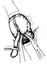
(3) Locating the appendix: Use gauze pads to push the small intestine medially, first locate the cecum, then trace along the three colonic bands to the apex of the cecum to find the appendix. If the appendix is still not found, consider the possibility of a retrocecal appendix, then incise the lateral posterior peritoneum and invert the cecum to locate the appendix. Once the appendix is found, use an appendix clamp or hemostatic forceps to grasp the mesoappendix and bring the appendix out through the incision for removal. If it cannot be brought out, the appendix should still be removed while strictly protecting the layers of the incision.
(4) Handling the mesoappendix: The appendicular artery is usually located at the free edge of the mesoappendix. When infection and inflammation worsen, the mesoappendix becomes fragile and easier to clamp off, so it is preferable to ligate the appendicular artery at the root of the appendix. If the mesoappendix is broad and thick, it should be ligated and cut in segments.
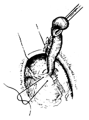
(5) Handling the root of the appendix: Gently clamp the root of the appendix 0.5cm from the cecum and ligate it with silk thread, then cut the appendix distal to the ligature. The stump is treated with iodine and alcohol, then buried into the cecal wall with a purse-string suture. The purse-string suture should not be too large to prevent leaving a dead space in the intestinal wall. Finally, reinforce with the mesoappendix or adjacent fatty connective tissue (Figure 1).
|
|
1. Cut the mesoappendix | 2. Perform a purse-string suture on the seromuscular layer of the cecal wall |
|
|
3. Resect the appendix at its base | 4. Tighten the purse-string suture and bury the stump into the cecal wall |
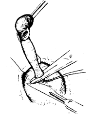
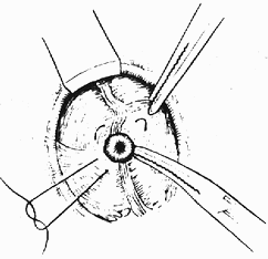
Figure 1 Appendectomy
(6) Appendectomy under special circumstances
1) If the appendix is located behind the peritoneum and is fixed by adhesions, making it difficult to remove by conventional methods, a retrograde resection should be performed. This involves first cutting the appendix at its base, burying the stump, and then sequentially cutting the mesoappendix to remove the entire appendix.
2) If the cecal wall is severely inflamed and edematous, making it impossible to bury the appendix stump into the purse-string suture as usual, the appendix can be cut at its base, and the stump can be buried using interrupted silk sutures in the seromuscular layer. If this is still not possible, the stump can be covered with the mesoappendix or nearby fatty connective tissue.
3) If the appendicitis is severely edematous, making the tissue fragile and prone to tearing, and the base cannot be clamped and ligated, a purse-string suture on the cecal wall can be used to invert and bury the unligated appendix stump into the cecal cavity, supplemented with interrupted silk sutures in the seromuscular layer.
I. Criteria for Cure:
1. The appendix is surgically removed, symptoms and signs disappear, the incision heals, and there are no complications.
2. After non-surgical treatment, symptoms and signs disappear.
II. Criteria for Improvement:
1. The appendix is not removed, symptoms are alleviated, and further surgical treatment is required.
2. After non-surgical treatment, symptoms and signs are alleviated.
1. Complications of acute appendicitis
(1) Abdominal abscess: An abscess formed around the appendix is called a periappendiceal abscess. However, abscesses can also form in other parts of the abdominal cavity, commonly in the pelvic cavity, subphrenic space, and interintestinal spaces. Clinical manifestations include symptoms of paralytic ileus such as abdominal distension and fullness, signs of peritoneal irritation, tender masses, and systemic infection and toxic symptoms. B-ultrasound can assist in diagnosis and localization. Once diagnosed, surgical incision and drainage should be performed promptly.
(2) Formation of internal and external fistulas: If a periappendiceal abscess is not drained in time, in some cases, the abscess may rupture into the small intestine or large intestine, or into the bladder, vagina, or abdominal wall, forming various internal or external fistulas; pus can be discharged from the fistula. X-ray barium examination can help understand the course and extent of the fistula, aiding in the selection of treatment methods such as extended drainage or fistula resection.
(3) Pylephlebitis: In acute appendicitis, infectious thrombi in the appendiceal vein can travel along the superior mesenteric vein to the portal vein, causing portal vein inflammation. Clinical manifestations include hepatomegaly and tenderness, jaundice, fear of cold, high fever, etc. If the condition worsens, it can lead to septic shock and sepsis, and delayed treatment can develop into bacterial liver abscess.
2. Complications of appendectomy
(1) Incision infection: This is the most common postoperative complication, with an incidence of less than 10% in the non-perforated group and more than 20% in the perforated group. It is mostly caused by contamination of the incision during surgery, retained hematoma and foreign bodies, and poor drainage. The infection site can be subcutaneous or outside the peritoneum. Clinical manifestations include elevated body temperature 2-3 days after surgery, local distending pain or throbbing pain at the incision site, with redness, swelling, and tenderness. Treatment involves cutting the sutures, enlarging the incision, draining the pus, removing foreign bodies, and ensuring adequate drainage.
(2) Peritonitis, abdominal abscess: Mostly caused by insecure ligation of the appendiceal stump and suture detachment. Clinical manifestations include persistent postoperative fever, abdominal pain, abdominal distension and fullness, and worsening systemic toxic symptoms. Treatment should follow the principles of treating peritonitis.
(3) Hemorrhage: Loosening of the ligature of the mesoappendix can cause massive intra-abdominal bleeding, presenting with abdominal pain, abdominal distension and fullness, hemorrhagic shock, etc. If the ligature of the appendiceal stump loosens and the purse-string suture is tight, bleeding can flow into the cecum, causing massive lower gastrointestinal bleeding. Both situations require immediate blood transfusion and fluid replacement, and emergency reoperation to stop the bleeding.
(4) Fecal fistula: There are various reasons for postoperative fecal fistula, such as fragile stump, ligature detachment; injury to the cecal wall; pre-existing conditions like subcutaneous nodules or cancer in the cecum; hard drainage material compressing the cecal wall causing necrosis, etc. Generally, when a fecal fistula forms, the inflammation is mostly localized, so diffuse peritonitis does not occur. The formed fecal fistula is located in the colon and does not cause water and electrolyte disturbances or nutritional disorders. Generally, after non-surgical supportive treatment, the fistula can close and heal on its own. If it does not heal for a long time, a biopsy of the fistula and X-ray fistulography can be performed to determine the nature and extent of the lesion, facilitating reoperation to remove the fistula.
(5) Stump appendicitis: If the stump is too long (more than 1 cm) during appendectomy, the stump is prone to recurrent inflammation after surgery, still presenting symptoms of appendicitis. Further X-ray barium examination should be performed to confirm the diagnosis. If the symptoms are severe, reoperation to remove the appendiceal stump is advisable.
(6) Adhesive intestinal obstruction: Due to surgical injury or factors such as pus around the appendix, some patients develop adhesive intestinal obstruction after surgery, especially after perforation, with an incidence of about 5%. Most cases can be effectively treated non-surgically, but severe cases require surgical treatment.
The clinical misdiagnosis rate of acute appendicitis is quite high, with domestic statistics showing 4-5%, and foreign reports indicating rates as high as 30%. There are many diseases that need to be differentiated from acute appendicitis, with the following dozen being the most significant.
I. Differentiation from medical acute abdomen:
1. Right lower lobe pneumonia and pleurisy: Inflammatory changes in the right lower lung and thoracic cavity can reflexively cause right lower abdominal pain, sometimes misdiagnosed as acute appendicitis. However, pneumonia and pleurisy often present with obvious respiratory symptoms such as cough, sputum, and chest pain, and chest signs like changes in breath sounds and rales. Abdominal signs are not obvious, and tenderness in the right lower abdomen is often absent. Chest X-rays can confirm the diagnosis.
2. Acute mesenteric lymphadenitis: More common in children, often secondary to upper respiratory infections. Due to widespread swelling of the small intestine mesenteric lymph nodes, especially at the end of the ileum, it can clinically present with right lower abdominal pain and tenderness, similar to acute appendicitis. However, this condition is accompanied by high fever, and the abdominal pain and tenderness are more widespread, sometimes palpable enlarged lymph nodes.
3. Regional ileitis: The lesion mainly occurs at the end of the ileum, a nonspecific inflammation, more common in young adults aged 20-30. In the acute phase, the affected intestinal segment becomes congested, edematous, and exudative, irritating the parietal peritoneum of the right lower abdomen, causing abdominal pain and tenderness similar to acute appendicitis. The pain is localized to the ileum without the characteristic of migratory abdominal pain, and abdominal signs are more widespread, sometimes palpable enlarged intestines. Additionally, patients may have diarrhea, and stool examination shows significant abnormal components.
II. Differentiation from gynecological acute abdomen:
1. Right tubal pregnancy: After rupture of a right ectopic pregnancy, intra-abdominal bleeding irritates the parietal peritoneum of the right lower abdomen, presenting clinical features similar to acute appendicitis. However, ectopic pregnancy often has a history of missed periods and early pregnancy, and may present with vaginal bleeding before onset. Patients may feel swelling in the perineum and anus after abdominal pain, along with signs of internal bleeding and hemorrhagic shock. Gynecological examination may reveal blood in the vagina, a slightly enlarged and tender uterus, a swollen right adnexa, and positive signs like blood on culdocentesis.
2. Ovarian cyst torsion: Torsion of the right ovarian cyst pedicle leads to circulatory disturbance, necrosis, and hemorrhagic exudation, causing inflammation in the right abdomen similar to appendicitis. However, this condition often has a history of pelvic mass, sudden onset, and presents with paroxysmal colicky pain, possibly accompanied by grade I shock symptoms. Gynecological examination can reveal a cystic mass with tenderness, and abdominal ultrasound confirms the presence of a cystic mass in the right lower abdomen.
3. Ovarian follicular rupture: More common in unmarried young women, often occurring two weeks after menstruation, causing right lower abdominal pain due to intra-abdominal bleeding. Local signs in the right lower abdomen are mild, and diagnostic peritoneal puncture can yield hemorrhagic exudate.
4. Acute adnexitis: Acute inflammation of the right fallopian tube can cause symptoms and signs similar to acute appendicitis. However, salpingitis is more common in married women with a history of excessive leucorrhea, often occurring before menstruation. Although there is right lower abdominal pain, it lacks the typical migratory nature, and the abdominal tenderness is lower, almost near the pubic area. Gynecological examination may reveal purulent vaginal discharge, significant tenderness on both sides of the uterus, and a tender mass in the right adnexa.
III. Differentiation from surgical acute abdomen:
1. Acute perforation of peptic ulcer: After perforation of a peptic ulcer, part of the gastric contents flow along the right paracolic gutter into the right iliac fossa, causing acute inflammation in the right lower abdomen, which can be mistaken for acute appendicitis. However, this condition often has a history of chronic ulcers, is triggered by overeating, and presents with sudden and severe abdominal pain. Physical examination reveals a board-like abdominal wall, with the most obvious peritoneal irritation signs below the xiphoid. Abdominal X-ray may show free gas under the diaphragm, and diagnostic peritoneal puncture can yield upper gastrointestinal fluid.
2. Acute cholecystitis and cholelithiasis: Acute cholecystitis sometimes needs to be differentiated from high appendicitis. The former often has a history of biliary colicky pain, accompanied by radiating pain to the right shoulder and back; while the latter is characterized by migratory abdominal pain. During examination, acute cholecystitis may present with a positive Murphy's sign, and even an enlarged gallbladder may be palpable. Emergency abdominal ultrasound can show gallbladder enlargement and acoustic shadows of stones.
3. Acute Meckel's diverticulitis: Meckel's diverticulum is a congenital malformation, mainly located at the end of the ileum, close to the appendix. When the diverticulum becomes acutely inflamed, the clinical symptoms are very similar to acute appendicitis, making it difficult to differentiate before surgery. Therefore, when the clinical diagnosis is appendicitis but the appendix appears normal during surgery, the terminal ileum should be carefully examined up to 1 meter to avoid missing an inflamed diverticulum that could lead to fistula disease.
4. Right ureteral stone: When a ureteral stone moves downward, it can cause pain in the lower right abdomen, which can sometimes be confused with appendicitis. However, the onset of a ureteral stone is characterized by severe colicky pain, which is unbearable and radiates along the ureter to the external genitalia and the inner thigh. On abdominal examination, tenderness and muscle tension in the lower right abdomen are not very obvious. An abdominal X-ray may sometimes reveal a positive stone in the urinary system, and a urine test may show a large number of red blood cells.


