| disease | Adult Umbilical Hernia |
| alias | Umbilical Hernia |
A hernia protruding from the umbilicus is called an umbilical hernia. Clinically, it is divided into two types: infant umbilical hernia and adult umbilical hernia.
bubble_chart Etiology
Umbilical hernias in adults are commonly seen in individuals with weak abdominal walls, such as those who are obese, middle-aged or elderly, and multiparous women. They are also frequently observed in patients with chronic conditions that increase intra-abdominal pressure. The hernial contents often include the greater omentum, followed by the transverse colon and small intestine.
bubble_chart Clinical Manifestations
The main clinical manifestation is a round hernial protrusion at the umbilicus when standing, coughing, or straining, which disappears when lying flat. After the hernia is reduced, the edges of the hernial ring can be palpated. If a significant amount of omentum and intestine protrudes, there may be dull pain and abdominal discomfort. Generally, smaller umbilical hernias may be asymptomatic. In adults, the edges of the hernial ring in umbilical hernias are tougher, less elastic, and non-expandable, with a higher incidence of incarceration and strangulation compared to infant umbilical hernias. The clinical presentation includes sudden severe pain, and if the contents involve the intestine, mechanical intestinal obstruction may occur.
bubble_chart Treatment Measures
Adult umbilical hernias cannot heal on their own and are prone to incarceration and strangulation, thus requiring surgical treatment. However, surgery is contraindicated in cases secondary to cirrhosis with ascites, or in elderly patients with severe cardiac or pulmonary diseases who cannot tolerate surgery. If incarceration or strangulation occurs, emergency surgery should still be performed.
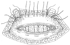
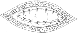
Make a transverse fusiform incision, incise the anterior rectus sheath, and transect the neck of the hernia sac at the hernia ring. Separate adhesions, excise the hernia sac wall and its outer covering, reduce the hernia contents, and perform high ligation of the hernia sac or suture the edges of the abdominal membrane. Mobilize the transversus abdominis membrane and rectus sheath around the hernia ring, and suture them in layers transversely. If the hernia ring is large, overlapping reinforcement sutures can be performed (Figure 1). It is worth noting that about 70% of adult umbilical hernias may be accompanied by rectus abdominis diastasis. For such umbilical hernias, a longitudinal incision is preferred to facilitate simultaneous repair and suturing of the separated rectus abdominis.
(1) After mobilizing the upper and lower leaves of the transversely incised rectus sheath, suture them

(2) Suture the edge of the lower leaf to the deep surface of the upper leaf

(3) Then overlap and suture the edge of the upper leaf to the superficial surface of the lower leaf
Figure 1 Umbilical hernia repair
In a few cases of large umbilical hernias, reduction may affect venous return to the heart, and elevation of the diaphragm may interfere with ventilation and oxygenation, particularly in elderly patients, requiring close monitoring of cardiac and pulmonary function.


