| disease | Primary Non-accommodative Esotropia |
| alias | Congenital Esotropia, Congenital Esotropia |
Congenital esotropia typically manifests as inward deviation of the eyes at birth or within a few days after birth. However, since parents rarely seek medical attention for newborns, clinically diagnosed cases of congenital esotropia are uncommon, and most cases involve early-onset strabismus detected shortly after birth. Parents often struggle to accurately and objectively assess the alignment of their infant's eyes within the first year of life, sometimes mistaking the instability of parallel visual axes for congenital esotropia. Additionally, during infancy, incomplete nasal development often leads to epicanthal folds and pseudostrabismus, which can further complicate the diagnosis. Some cases of acquired accommodative esotropia may also emerge during this period, all of which can contribute to diagnostic confusion.
bubble_chart Clinical Manifestations
The clinical features of congenital strabismus include a large deviation angle, stable strabismus angle, and accompanying abnormal eye movements. Congenital esotropia can occur in otherwise normal children, as well as in infants with cerebral palsy or hydrocephalus. In children with brain injuries, the deviation angle often varies with age and may disappear as they grow older. Occasionally, between 6 months to 1 year of age, esotropia may change into exotropia.
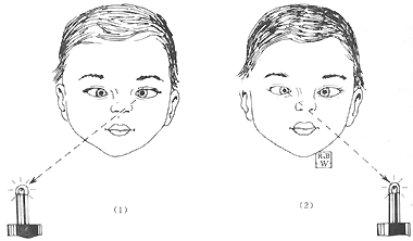
1. Most patients with congenital esotropia exhibit alternate fixation in the primary position, with equal visual acuity in both eyes. They show cross-fixation when looking to either side (Figure 1), meaning the left eye fixes when looking to the right, and the right eye fixes when looking to the left. A minority of patients lack alternate fixation, and the deviated eye may develop amblyopia, with an incidence rate of approximately 40%. The amblyopia is often severe and accompanied by eccentric fixation.

Figure 1 Cross-fixation
(1) Left eye fixes when looking to the right (2) Right eye fixes when looking to the left
2. The angle of deviation is large, typically exceeding 30△, with about 50% of patients exceeding 50△. The angle remains consistent for both distance and near vision, is stable, and unaffected by accommodation. Occasionally, the angle may change significantly over a few months. It should be noted that affected children often cannot abduct both eyes, but this is not due to bilateral abducens nerve palsy; rather, it is secondary to cross-fixation. Another scenario is when children with congenital esotropia have a large deviation and amblyopia but no cross-fixation, which may lead to misdiagnosing the eccentrically fixing eye as unilateral abducens nerve palsy. In reality, congenital unilateral or bilateral abducens nerve palsy is rare.
Congenital esotropia should also be differentiated from Duane retraction syndrome, Mobius syndrome, and abducens nerve palsy. The differentiation methods are as follows: ① Fix the child’s head in an upright position and gently rotate it horizontally, both quickly and slowly, to stimulate the labyrinth, particularly the horizontal semicircular canals. A subtle abduction movement may momentarily appear and can be detected with close observation. ② In congenital esotropia with cross-fixation, patching one eye for several days may induce abduction in the other eye. ③ Forced duction testing under general anesthesia shows normal results in congenital esotropia with cross-fixation, with no passive resistance during abduction. As anesthesia deepens, the esotropia may disappear and even turn into exotropia.
3. Frequent association with vertical deviations: By the age of 2–3 years, children with congenital esotropia may develop dissociated vertical deviation (DVD), characterized by hypertropia and excyclotorsion of the non-fixing eye and hypotropia and incyclotorsion of the fixing eye. About 78% of patients exhibit overaction of the inferior oblique muscle. Ocular tremor, either torsional or horizontal, may also be observed. The tremor is sometimes latent, appearing only after covering one eye or manifesting as reduced nystagmus in adduction and increased nystagmus in abduction.
4. Cycloplegic refraction reveals that 90% of cases have grade I or grade II hyperopia, with similar refractive errors in both eyes. Astigmatism or myopia may also be present.5. The AC/A ratio is normal.
6. ① Measuring the angle of deviation: Since prism-cover testing is difficult in infants, the Hirschberg and Krimsky methods are commonly used. The child is asked to fixate on a light, and the angle is determined by the prism power required to center the corneal light reflex. ② Cycloplegic refraction. ③ Sensory testing in older children.
bubble_chart Treatment Measures
1. Hyperopia greater than +2.00D should be corrected.
2. For patients with amblyopia, occlusion therapy can be used. Alternate occlusion is effective for a small number of amblyopia patients but ineffective in preventing suppression and abnormal retinal correspondence because, during infancy, only orthophoria or esotropia within 10△ can produce binocular single vision. Therefore, it is incorrect to use alternate occlusion for several years in early congenital esotropia patients and then train fusion before performing surgery.
3. For cases less than 1.50D, strong miotics can be used once daily for 2–3 weeks. When the child is ≥6 months old and capable of alternate fixation, surgery may be considered.
4. Surgical treatment: The primary treatment for congenital esotropia is surgical correction of ocular alignment, with debate focusing on when and how to perform the surgery. Parks, Taylor, and Costenbader advocate surgery within 6–12 months, with Parks suggesting that surgery during 6–12 months offers a better chance of postoperative fusion recovery compared to 12–18 months. Von Noorden, Jampolsky, and others, based on research, have demonstrated that congenital esotropia corrected after 1 year of age can achieve binocular fusion. They argue that correcting alignment before age 2 does not yield a higher rate of central fusion than surgery performed at 12–18 months. Additionally, their research further shows that postoperative optical correction—using prisms or minus lenses to address residual deviation—significantly increases the rate of central fusion: about 53% of children achieve central fusion with optical correction, compared to only 6% with surgery alone. Moreover, children under 1 year present challenges in examination, diagnosis, and precise measurement. Inadequate preoperative preparation increases the risk of overcorrection or undercorrection, leading most ophthalmologists to recommend the optimal timing for surgery as 12–18 months of age, with the latest being no later than 2 years. Parks notes that even with parallel visual axes postoperatively, congenital esotropia patients often achieve only peripheral fusion rather than central fusion, termed the monofixation syndrome. This is crucial because it helps prevent recurrence of esotropia or progression to exotropia.
Surgical techniques include bilateral medial rectus recession or medial rectus recession combined with lateral rectus resection. For deviations of 50–75△, three horizontal muscles may be operated on; for deviations of 70–90△, all four horizontal rectus muscles may be addressed. Overaction of the inferior oblique muscle often requires inferior oblique recession. For children under 4 years, congenital esotropia should be corrected to within 10△, while older children may tolerate undercorrection of up to 15△ to preserve the chance of binocular vision restoration. In older children, the goal is cosmetic alignment; if binocular vision is absent, exotropia may develop years postoperatively. Residual esotropia of 15△ can delay exotropia onset. The occurrence of exotropia 10–30 years postoperatively is not a reason to reject surgery, as exotropia can be corrected with additional surgery. To achieve binocular single vision in congenital esotropia, early surgery is essential to maintain orthophoria. After treatment, accommodative esotropia and amblyopia may still develop, so close follow-up and appropriate therapy are necessary for at least 5 years postoperatively.


