| disease | Aortic Valve Insufficiency |
| alias | Aortic Insufficlency |
Aortic insufficiency can be caused by lesions in the aortic valve, valve annulus, and ascending aorta. It is more common in male patients, accounting for about 75%. Female patients often have concurrent mitral valve disease. Among chronic cases, valve leaflet damage due to wind-dampness heat is the most common cause, accounting for two-thirds of all patients with aortic insufficiency.
bubble_chart Etiology
Acute aortic valve insufficiency is commonly seen in infective endocarditis, where infection damages the valve, causing leaflet perforation, or where vegetations prevent complete leaflet coaptation, or where post-inflammatory healing leads to scarring and contracture, or where leaflet degeneration and prolapse occur, all of which can result in aortic regurgitation. Trauma-induced aortic valve insufficiency is relatively rare and may occur after aortic valve stenosis separation or valve replacement surgery, or it may result from non-penetrating ascending aortic tears caused by trauma. Retrograde aortic dissection involving the aortic valve annulus can also lead to acute or chronic aortic valve insufficiency.
The main pathophysiological changes of aortic valve insufficiency are due to the left ventricular pressure during diastole being significantly lower than the aortic pressure, causing a large amount of blood to regurgitate back into the left ventricle. This increases the diastolic load on the left ventricle (normal left atrial return and abnormal aortic regurgitation), leading to a gradual increase in left ventricular end-diastolic volume, while the end-diastolic pressure may remain normal. Due to the decreased resistance in the aorta from blood regurgitation, the left ventricular stroke volume increases early in systole, and the ejection fraction remains normal. As the condition progresses, the regurgitant volume increases, potentially reaching 80% of the stroke volume. The left ventricle further dilates, myocardial hypertrophy occurs, and both the end-diastolic volume and pressure of the left ventricle rise significantly, along with a marked increase in systolic pressure. When left ventricular contraction weakens, the stroke volume decreases. In the early stages, the resting grade I is reduced and fails to increase during exercise. In the advanced stage, the left ventricular end-diastolic pressure rises, leading to increased pressure in the left atrium, pulmonary veins, and pulmonary capillaries, followed by dilation and congestion. When aortic valve regurgitation is significant, the aortic diastolic pressure drops markedly, reducing coronary perfusion pressure. Myocardial blood supply decreases, further weakening myocardial contractility.
In acute aortic valve insufficiency, the left ventricle suddenly receives a large volume of regurgitant blood, but the stroke volume cannot increase accordingly. The left ventricular end-diastolic pressure rises rapidly and significantly, potentially causing acute left heart dysfunction. The elevated end-diastolic pressure reduces the pressure gradient between coronary perfusion pressure and left ventricular cavity pressure, leading to subendocardial myocardial ischemia and weakened myocardial contractility. These factors can cause a sharp decline in stroke volume, a rapid rise in left atrial and pulmonary venous pressure, and result in acute pulmonary edema. During this time, sympathetic nervous activity increases significantly, raising the heart rate and peripheral vascular resistance. The decrease in diastolic pressure may not be significant, and the pulse pressure remains small.bubble_chart Pathological Changes
The pathological changes mainly involve inflammation and fibrosis causing the valve leaflets to become stiff, shortened, and deformed, leading to abnormalities in leaflet opening during systole and closure during diastole. Most patients also have aortic valve stenosis. Aortic valve insufficiency can also be seen in congenital malformations such as bicuspid aortic valve, fenestrated aortic valve, ventricular septal defect with aortic valve prolapse, etc., as well as connective tissue diseases like systemic lupus erythematosus and rheumatoid arthritis. The mucoid degeneration of the valve leaflets that causes mitral valve prolapse syndrome can also affect the aortic valve, resulting in aortic valve insufficiency. Diseases of the ascending aorta can lead to dilation of the aortic root, causing enlargement of the aortic valve annulus and incomplete closure of the normal aortic valve during diastole, resulting in aortic regurgitation. Common causes include Marfan syndrome, ascending aortic atherosclerosis, aortic sinus aneurysm, syphilitic aortitis, cystic medial necrosis of the ascending aorta, severe hypertension, and idiopathic aortic dilation.
bubble_chart Clinical Manifestations(1) Symptoms Typically, patients with aortic regurgitation remain asymptomatic for a prolonged period, even in cases of significant aortic regurgitation, where noticeable symptoms may take 10 to 15 years to manifest. However, once heart failure develops, progression is rapid.
1. Palpitation Discomfort from heart palpitations may be the earliest complaint, caused by the pronounced enlargement of the left ventricle and intensified apical pulsations, particularly noticeable when lying on the left side or in a prone position. Emotional stress or physical exertion leading to tachycardia or ventricular premature beats can exacerbate the sensation of palpitation. Due to a significant increase in pulse pressure, patients often experience intense pulsations throughout the body, especially in the head and neck.
2. Dyspnea Exertional dyspnea is the earliest symptom, indicating reduced cardiac reserve. As the condition progresses, orthopnea and paroxysmal nocturnal dyspnea may occur.
3. Chest Pain Angina is less common in aortic regurgitation compared to aortic stenosis. Chest pain may result from excessive stretching of the ascending aorta during left ventricular ejection or significant cardiac enlargement, with myocardial ischemia also playing a role. Angina can occur during activity or at rest, lasting for extended periods and responding poorly to nitroglycerin. Nocturnal angina attacks may be due to a further drop in diastolic pressure during rest, reducing coronary blood flow. Some patients report abdominal pain, possibly related to visceral ischemia.
4. Syncope Dizziness or vertigo may occur with rapid postural changes, though syncope is rare.
5. Other Symptoms Fatigue and markedly reduced exercise tolerance. Excessive sweating, particularly during episodes of paroxysmal nocturnal dyspnea or nocturnal angina. Hemoptysis and embolic events are uncommon. In advanced stages, right heart failure may lead to congestive hepatomegaly with tenderness, ankle edema, pleural effusion, or ascites.In acute aortic regurgitation, the sudden increase in left ventricular volume load and wall tension, along with left ventricular dilation, can rapidly lead to acute left heart failure or pulmonary edema.
(2) Signs
1. Cardiac Auscultation A diastolic murmur is heard at the aortic valve area, characterized as a high-pitched, decrescendo blowing sound, most prominent when the patient sits forward at the end of expiration. The loudest area depends on the degree of ascending aortic dilation: in rheumatic cases with mild aortic dilation, the murmur is loudest at the third left intercostal space along the sternal border, radiating to the apex; in Marfan syndrome or syphilitic heart disease, where the ascending aorta or aortic annulus may be severely dilated, the murmur is loudest at the second right intercostal space. Generally, the more severe the aortic regurgitation, the longer and louder the murmur. In grade I regurgitation, the murmur is soft and limited to early diastole, audible only when the patient sits forward at the end of expiration. In more severe cases, the murmur may be holodiastolic and harsh. In grade III or acute aortic regurgitation, the murmur duration may shorten due to elevated left ventricular end-diastolic pressure equaling aortic diastolic pressure. A musical murmur suggests partial valve leaflet eversion, tearing, or perforation. Aortic dissection may also produce a musical murmur, possibly due to diastolic prolapse of the proximal aortic intima through the aortic valve or blood flow within the dissected aortic lumen.
In cases of significant aortic regurgitation, a systolic ejection murmur—soft, short, and high-pitched—is often heard at the base of the heart in the aortic valve area, radiating to the neck and suprasternal notch. This is caused by the large stroke volume passing through the deformed aortic valve membrane, rather than by organic aortic stenosis. A soft, low-pitched rumbling diastolic or presystolic murmur, known as the Austin-Flint murmur, is frequently audible at the apex. This occurs due to massive aortic regurgitation, which impacts the anterior leaflet of the mitral valve, impeding its opening and causing vibrations, leading to relative mitral stenosis. Additionally, the regurgitant aortic flow collides and mixes with the left atrial return flow, generating vortices. This murmur intensifies with forceful handgrip and diminishes with amyl nitrite inhalation. When the left ventricle is significantly dilated, functional mitral regurgitation may occur due to outward displacement of the papillary muscles, resulting in a holosystolic blowing murmur at the apex that radiates to the left axilla.
When the aortic valve membrane activity is poor or regurgitation is severe, the aortic valve's second heart sound weakens or disappears; a third heart sound is often audible, indicating left ventricular dysfunction; a fourth heart sound may be heard when left atrial compensatory contraction is enhanced. Due to the significant increase in stroke volume during systole, the sudden expansion of the aortic valve can produce a loud early systolic ejection sound.
In acute severe aortic valve regurgitation, the diastolic murmur is soft and short; the first heart sound weakens or disappears, and a third heart sound may be audible; pulse pressure may be nearly normal.
2. Other signs: The face appears pale, the apical impulse shifts downward and to the left with a wide range and exhibits a forceful heaving motion. The cardiac dullness border expands downward and to the left. A systolic thrill can be palpated at the aortic valve area and transmitted to the neck; a diastolic thrill may be palpated at the lower left sternal border. Carotid artery pulsations are markedly enhanced and exhibit a double beat. Systolic blood pressure is normal or slightly elevated, while diastolic pressure is significantly reduced, resulting in a markedly widened pulse pressure. Peripheral vascular signs may appear: water-hammer pulse (Corrigan's pulse), capillary pulsation sign (Quincke's sign), pistol-shot sound over the femoral artery (Traube's sign), systolic and diastolic double murmur over the femoral artery (Duroziez's sign), and rhythmic nodding of the head with each heartbeat (de Musset's sign). With pulmonary hypertension and right heart failure, distended neck veins, hepatomegaly, and lower limb edema may be observed.
bubble_chart Auxiliary Examination
1. X-ray examination shows significant enlargement of the left ventricle, dilation of the ascending aorta and aortic root, presenting as an "aortic-type heart." Fluoroscopy reveals markedly enhanced aortic pulsation, which, in coordination with left ventricular pulsation, exhibits a "rocking chair-like" motion. The left atrium may also be enlarged. In cases of pulmonary hypertension or right heart failure, the right ventricle enlarges. Pulmonary venous congestion and interstitial edema may be visible. Calcification of the aortic valve leaflets and ascending aorta is often present. Aortography can assess the severity of aortic regurgitation. If the contrast reflux into the left ventricle is denser than that in the ascending aorta, it indicates grade III regurgitation; if the reflux is limited to the subvalvular area or appears as a linear jet, it is classified as grade I regurgitation.
2. Electrocardiogram (ECG) findings in grade I aortic regurgitation may be normal. Severe cases may exhibit left ventricular hypertrophy and strain, with left axis deviation. Deep Q waves, ST-segment depression, and T-wave inversion are seen in leads I, aVL, and V5–6. Advanced stages may show left atrial enlargement. Bundle branch block may also be observed.
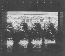
3. Echocardiography reveals dilation of the left ventricular cavity, left ventricular outflow tract, and ascending aortic root. When myocardial contractility is compensatory, the systolic motion amplitude of the left ventricular posterior wall increases; the rate and amplitude of ventricular wall motion remain normal or elevated. A characteristic feature of aortic regurgitation is the rapid, high-frequency vibration of the anterior mitral leaflet during diastole (Figure 1). Two-dimensional echocardiography may show thickening of the aortic valve with poor coaptation during diastole. Doppler ultrasound detects diastolic turbulence below the aortic valve, which is highly sensitive for identifying aortic regurgitation and assessing its severity. Echocardiography is also valuable for evaluating left ventricular function in aortic regurgitation and aids in determining the etiology, such as bicuspid aortic valve, valve prolapse, rupture, vegetation formation, or ascending aortic dissection.

Figure 1: M-mode echocardiogram of aortic regurgitation
showing fine fluttering waves of the anterior mitral leaflet during diastole (downward arrow).
4. Radionuclide imaging demonstrates left ventricular enlargement and increased end-diastolic volume. The left atrium may also be dilated. This method can measure left ventricular systolic function and is useful for follow-up assessments.
The clinical diagnosis is primarily based on the typical diastolic murmur and left ventricular enlargement, with echocardiography providing definitive confirmation. The disease cause can be determined based on medical history and other findings.
bubble_chart Treatment Measures
(1) Medical treatment: Avoid excessive physical labor and strenuous exercise, limit sodium intake, and use Rehmannia-based medications, diuretics, and vasodilators, especially angiotensin-converting enzyme inhibitors, to help prevent deterioration of cardiac function. Rehmannia-based medications can also be used in patients without symptoms of heart failure but with severe aortic regurgitation and significant left ventricular enlargement. Active prevention and treatment of arrhythmias and infections are essential. Syphilitic aortitis should be treated with a full course of penicillin therapy, while rheumatic heart disease requires active prevention of streptococcal infections, rheumatic activity, and infective endocarditis.
(2) Surgical treatment: Artificial valve replacement is the primary method for treating aortic regurgitation and should be performed before symptoms of heart failure appear. However, since patients typically show no obvious symptoms before myocardial decompensation occurs, surgery is not urgently needed in asymptomatic patients with normal left ventricular function. Close follow-up is recommended, with echocardiography repeated at least every six months. Surgery should be performed once symptoms, left ventricular dysfunction, or significant cardiac enlargement occur.
1. Valve repair: Rarely used and usually cannot completely eliminate aortic regurgitation. It is only suitable for cases of infective endocarditis with aortic valve vegetations or perforations, or tears between the aortic valve and its annulus. Aortic regurgitation caused by dilation of the annulus due to ascending aortic aneurysm may be treated with annuloplasty.
2. Artificial valve replacement: Rheumatic and most other causes of aortic regurgitation are best treated with valve replacement. Both mechanical and biological valves can be used. Surgical risk and late mortality depend on the stage of aortic regurgitation and the patient's cardiac function at the time of surgery. Patients with significant cardiac enlargement and long-term left ventricular dysfunction have a surgical mortality rate of about 10% and a late mortality rate of about 5% per year. Nevertheless, due to the poor prognosis of medical treatment, surgical intervention should be considered even in cases of left ventricular failure.
(3) Treatment of acute aortic regurgitation: Severe acute aortic regurgitation rapidly leads to acute left ventricular dysfunction, pulmonary edema, and hypotension, which are highly fatal. Therefore, surgical treatment should be performed as early as possible alongside aggressive medical therapy to save the patient's life. Preoperatively, positive inotropic agents such as dopamine or dobutamine and vasodilators such as nitroprusside should be administered intravenously to maintain cardiac function and blood pressure.
Congestive heart failure is common and is the main cause of death in patients with regurgitant valvular disease. Once symptoms of cardiac dysfunction appear, death often occurs within 2 to 3 years. Infective endocarditis can also occur, while embolism is rare.
Aortic valve insufficiency should be differentiated from the following diseases:
(1) Pulmonary valve insufficiency. This condition is often caused by pulmonary hypertension. In this case, the jugular pulse is normal, the second heart sound at the pulmonary valve area is accentuated, the diastolic murmur at the left sternal border intensifies during inspiration, and there is no change when forcefully clenching the fist. The electrocardiogram shows hypertrophy of the right atrium and right ventricle, and X-ray examination reveals prominence of the pulmonary artery trunk. It is commonly seen in mitral stenosis and can also occur in atrial septal defects.
(2) Rupture of an aortic sinus aneurysm. The rupture often extends into the right heart, producing a continuous murmur at the lower left sternal border. However, sometimes the murmur is to-and-fro, resembling aortic valve insufficiency with a concomitant systolic murmur. However, there is sudden onset of chest pain and progressive right heart failure. Aortic angiography and echocardiography can confirm the diagnosis.
(3) Coronary arteriovenous fistula. This often causes a continuous murmur, but a diastolic murmur may also be heard at the aortic valve area, or the diastolic component of the murmur may be louder. However, the electrocardiogram and X-ray examination are usually normal. Aortic angiography may reveal a communication between the aorta and the coronary sinus, right atrium, ventricle, or main pulmonary artery.


