| disease | Chronic Suppurative Otitis Media |
Chronic suppurative otitis media is defined as persistent ear discharge lasting more than 1 to 2 months due to improper treatment of acute suppurative otitis media, highly virulent bacteria, weakened body resistance, or complications such as mastoiditis. This condition has a relatively high incidence rate. A recent domestic survey of over a thousand primary school students revealed an incidence rate of 0.5% to 4.3%, while surveys of farmers in Shandong, Henan, and Guizhou provinces showed an incidence rate of 1.6%. In the UK, the incidence rate among primary school students was found to be 0.9%. The majority of affected individuals are young adults, with rare occurrences after the age of 40.
bubble_chart Etiology
1. Delayed treatment during the acute phase and improper medication, etc.
2. Poor mastoid development, making it difficult for lesions to dissipate after onset.
3. Secondary to acute pestilential diseases such as scarlet fever, measles, and pneumonia, acute necrosis of the middle ear mucosa occurs, with inflammation invading the tympanic antrum and mastoid. This is especially challenging when caused by highly drug-resistant infections like Proteus or Pseudomonas aeruginosa, making treatment extremely difficult.
4. Chronic nasal and pharyngeal conditions such as sinusitis, tonsillitis, and adenoid hypertrophy allow inflammatory secretions to easily enter the Eustachian tube, while the lesions obstruct drainage at the pharyngeal orifice.
5. Chronic systemic diseases like anemia, diabetes, pulmonary subcutaneous nodules, and nephritis weaken the body's resistance.
6. Presence of allergic conditions, such as allergic edema and exudation in the upper respiratory mucosa, affecting the Eustachian tube and middle ear.
7. Development of cholesteatoma in the epitympanum, ossicular necrosis, or destruction of the lateral wall of the tympanic cavity. {|106|}bubble_chart Pathological Changes
According to the severity and risk level of the lesions, they are divided into three types.
(1) Simple Type Also known as the tubotympanic type, it is the most common, with lesions mainly confined to the tympanic cavity. The normal Eustachian tube and anterior tympanum are covered by ciliated columnar epithelium, containing glands, while the posterior tympanum, tympanic antrum, and mastoid are lined with cuboidal epithelium. The ossicles, muscles, ligaments, and nerves in the tympanic cavity are all surrounded by mucous membrane, forming many folds and shallow pockets. Generally, if the mucous membrane becomes infected and inflamed, timely treatment can lead to perforation of the tympanic membrane, ensuring proper drainage and rapid resolution of the inflammation. Otherwise, the lesions in the shallow pockets may expand, and the mucous membrane lesions may become irreversible. Although the pus discharge may not be excessive, it can persist for a long time or recur shortly after temporary relief. The mastoid is usually well-pneumatized and unaffected.
(2) Necrotic Type Also known as the osteitic type. The mucous membrane and tissues are extensively destroyed, and hemorrhage and necrosis may occur in the ossicles, tympanic ring, tympanic antrum, and mastoid air cells. Perforations are particularly common in the flaccid part and the upper posterior tympanum. The pus is not abundant but has a strong foul odor, and granulation tissue and polyps are often seen obstructing drainage within the perforation. Severe hearing impairment may occur, and sometimes headache and vertigo may be present. The mastoid is often of the interstitial or sclerotic type.
(3) Cholesteatoma Type Also known as the dangerous type. Hyperplastic epithelial masses form in the tympanic cavity or tympanic antrum, surrounded by fibrous tissue and containing necrotic material, keratinized debris, and cholesterol crystals. Because it can erode and destroy bone, it exhibits malignant tumor-like properties, hence it was mistakenly referred to as cholesteatoma in the past, though it is not actually a tumor. The ear discharge is not copious but has an extremely foul odor, and white fragments or cheesy cholesteatomatous epithelial masses can be seen within the perforation. It may cause headache and dizziness. Due to extensive bone destruction, it is prone to intracranial and extracranial complications, hence it is called dangerous otitis media. The mastoid is usually of the sclerotic type (Table 1).Table 1 Differentiation of the Three Types of Chronic Suppurative Otitis Media
| Simple Type | Necrotic Type | Cholesteatoma Type | |
| Nature of Discharge | Mucoid or mucopurulent, not thick, white or pale yellow, mild odor | Purulent, not thick, yellow, sometimes bloody, foul-smelling | Purulent, scanty, very thick, with crusts, yellow, extremely foul odor, resembling rotten eggs |
| Duration of Discharge | Recurrent, intermittent | Continuous | Continuous or intermittent, very little discharge |
| Tympanic Membrane Perforation | Mostly small central perforation | Large central perforation or marginal perforation | Flaccid part or marginal perforation |
| Ossicular Chain | Mostly normal, tympanic mucous membrane edema | Ossicular destruction, granulation tissue and polyps in tympanum | Ossicular destruction, granulation tissue and cholesteatoma in tympanum |
| Deafness | Grade I conductive deafness | Grade II conductive deafness | Grade III conductive deafness or mixed deafness |
| Cholesteatoma | Absent | Rare | Common |
| Mastoid X-ray | Increased small room density, no bone destruction | Interstitial papillary osteomyelitis, with bone destruction | Sclerotic mastoid, often with well-defined round bone destruction |
Middle ear mucosal inflammation can induce cholesterol sebaceous cysts containing cholesterol crystals and cholesterol granulomas. The latter, although classified under choleatosis, is merely a granuloma and differs significantly from the epithelial accumulation masses of cholesterol sebaceous cysts. The key pathological distinctions between the two are as follows:
1. Cholesterol granuloma: Caused by eustachian tube obstruction, negative pressure in the tympanic cavity, exudation or formation of glue ear, capillary hemorrhage, and deposition of cholesterol crystals and hemosiderin on the epithelial surface. The tympanic membrane appears blue, and the mastoid air cells show mucosal edema. Microscopically, the typical manifestation is a cholesterol granuloma, with cholesterol crystals surrounded by foreign-body giant cells and an outer layer of fibrous granulation tissue. It is commonly seen in hemorrhagic and necrotic sexually transmitted diseases of the tympanic cavity and is not a precursor to cholesterol sebaceous cysts, nor is it related to their formation.
2. Cholesterol sebaceous cysts: There are two mechanisms of formation.
(1) Congenital cholesterol sebaceous cysts: Very rare. These are epithelial masses formed due to the proliferation of residual embryonic epithelial tissue in the middle ear under certain stimuli. They are usually located in the epitympanum and may occur without a history of otitis media. The tympanic membrane remains entirely normal until the cyst expands and perforates it, leading to secondary infection and pus discharge.
(2) Acquired cholesterol sebaceous cysts: These form due to excessive epithelial proliferation stimulated by localized suppurative otitis media, accounting for 30% of chronic otitis media cases. The exact cause remains debated. The currently accepted theories include: - The epithelial migration theory: The basal cells of the germinal layer of the external auditory canal skin possess unique proliferative potential. Under the stimulation of otitis media, these basal cells proliferate and invade the subepithelial connective tissue of the middle ear mucosa or form granulomas. Concurrently, subepithelial sclerosis leads to new bone formation, and the mass enlarges, eventually causing secondary perforation of the tympanic membrane. The keratinized layers of the epithelial mass slough off and necrotize, leading to secondary infection and the release of cholesterol and various chemical decay products. These physicochemical factors can erode and destroy surrounding bone, exposing adjacent meninges, nerves, and blood vessels, resulting in numerous intracranial and extracranial complications. Their tissue-destructive properties resemble tumors, leading Wendt (1873) to first name them cholesterol sebaceous cysts. In essence, they are not tumors, but the term has been used for so long that it awaits future correction. - Another view suggests that upper respiratory infections induce eustachian tube obstruction, creating negative pressure in the tympanic cavity. This causes inward invasion of the tympanic membrane's flaccid portion or the entrapment of posterior external auditory canal epithelium into the mastoid antrum, forming a pouch—the precursor to a cholesterol sebaceous cyst. This stage can persist for years. If accumulated keratin is promptly removed during this stage, the formation of a cholesterol sebaceous cyst can be prevented. Otherwise, once the accumulated epithelial mass becomes infected, it can rupture into the tympanic cavity, forming perforations in the flaccid or marginal regions and leading to cholesterol sebaceous cysts (Figure 1).
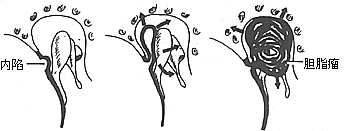
Figure 1: Schematic diagram of inward invasion of the flaccid portion forming a cholesterol sebaceous cyst pouch.
bubble_chart Clinical Manifestations
1. The nature and duration of pus discharge vary depending on the severity of the lesion. In mild cases, it is mucopurulent and intermittent, with alternating periods of improvement and worsening; in severe cases, it is persistent, presenting as thick yellow pus with a foul odor.
2. During acute episodes, symptoms may include headache, ear pain, dizziness, and fever. In severe cases, deviation of the mouth and meningitis may occur.
3. In the early stage, the tympanic membrane shows a central circular or reniform perforation. Occasionally, small perforations in the flaccid part or marginal area may be observed, often covered by purulent scabs with minimal discharge. If the scabs are not carefully removed, it can easily lead to misdiagnosis as fistula disease. {|102|}
Based on medical history and clinical manifestations, the diagnosis is not difficult. Granulation tissue from the external auditory canal and perforation sites should be sent for pathological examination to rule out tumors. Pus crusts and secretions must be removed to clearly observe whether there is a tympanic membrane perforation, which can be examined using an otoscope or Siegle's otoscope. If there is pus with a peculiar odor, a small amount can be taken and dissolved in chloroform or acetone. If it turns yellow-green, it suggests the formation of a cholesteatoma. A tuning fork and audiometer can be used to assess the nature and degree of deafness. Routine mastoid X-rays are usually taken in the Law or Meyer position to observe for bone destruction. If necessary, a mastoid CT scan can be performed to check for cholesteatoma-related bone destruction.
bubble_chart Treatment Measures
(1) Local Treatment
According to domestic bacterial culture of chronic suppurative otitis media, the predominant pathogens are Staphylococcus aureus and Haemophilus influenzae, with an increasing prevalence of penicillin-resistant Gram-positive bacteria. Conventional broad-spectrum antibiotics administered orally or intravenously often prove ineffective, particularly because the blood vessels beneath the middle ear and mastoid mucosa have become fibrotic due to scarring, preventing adequate drug concentrations from reaching the affected area. Moreover, antagonism may lead to bacterial drug resistance, making topical medication more advantageous. Pus cultures with antibiotic sensitivity tests should be performed to select effective drugs. Commonly used preparations and methods are similar to those for acute suppurative otitis media, but this approach is only suitable for Type I or II chronic otitis media. Before medication, purulent crusts in the external auditory canal must be removed. The patient should lie on their side with the affected ear facing upward. After instilling the medication, the air displacement method is employed by pressing the tragus. Ideally, a suction device should be used to thoroughly clean the area before forcing the medication into the tympanic cavity and mastoid air cells. Some cases of long-term purulent Type I otitis media can be cured within 1–2 months with regular and appropriate treatment. Otherwise, improper medication or failure to adhere to a daily instillation schedule will make it difficult to achieve a cure.
(2) Surgical Treatment
1. Chronic Simple and Osteitic Otitis Media
(1) Removal of Adjacent Infectious Foci: Hypertrophic nasal turbinates, nasal polyps, deviated nasal septum, and other conditions impairing nasal airflow should be surgically excised or corrected. Chronic sinusitis requires radical treatment, while chronic tonsillitis and adenoid hypertrophy should be removed, especially in children. Adenoid hypertrophy and inflammation are often the cause of persistent otitis media, and their removal frequently accelerates recovery.
(2) Tympanoplasty: To eradicate lesions and restore hearing, Wüllstein and Zöllner pioneered tympanoplasty in the 1950s, which has since been widely adopted. In 1956, Wüllstein classified tympanoplasty into five types: - **Type I (Myringoplasty)**: Suitable for cases without granulation, cholesteatoma, or bony pathology in the tympanic cavity. Successful repair of the tympanic membrane significantly improves hearing. - **Type II (Atticoantrostomy)**: Indicated for marginal or flaccid perforations of the tympanic membrane with granulation, cholesteatoma, or bony pathology. - **Type III (Columella Technique)**: For cases with mild to moderate disease, interrupted ossicular chain but intact stapes. Lesions are removed, and the residual tympanic membrane or graft is adhered to the stapes to reconstruct the tympanic cavity or ossicular chain, improving hearing. - **Type IV (Total Tympanoplasty and Small Cavity Technique)**: For complete ossicular destruction. After lesion removal, the residual tympanic membrane or graft is used to create a small cavity connecting the round window and Eustachian tube, enhancing sound transmission by increasing the impedance difference between the two windows. - **Type V (Small Cavity with Fenestration)**: For cases with missing ossicles and stapes fixation by granulation or scar tissue. In addition to creating a small cavity, a fenestration is made in the horizontal semicircular canal to allow sound waves to enter the inner ear through the new window, improving hearing (Figure 1).
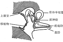
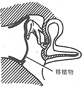

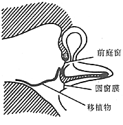
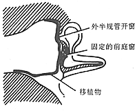
Figure 1 Wullstein’s Classification of Tympanoplasty
These five types are somewhat ambiguous in content, some involving surgeries of the mastoid antrum and mastoid process. The Hearing Preservation Committee of the American Academy of Ophthalmology and Otolaryngology proposed that the definition of tympanoplasty should be a surgery that eliminates diseases in the tympanic cavity and reconstructs the auditory mechanism, including only the repair of the tympanic membrane and ossicles, and not mastoid surgery. If the mastoid is involved, it should be labeled as tympanoplasty plus mastoid surgery. In China, types I, II, III, and IV of tympanoplasty are commonly used, while type V is rarely adopted. Typically, type I refers to tympanic membrane repair, and type VI refers to radical mastoidectomy.
2. Surgery for severe osteomyelitic and cholesteatomatous otitis media Due to conditions such as osteomyelitis, granulation tissue, and cholesteatoma, the primary goal is to remove the lesions to achieve a dry ear, with hearing improvement pursued when possible. For cases with cholesteatoma, complete removal of the lesions is essential to prevent intracranial or extracranial complications. Several representative surgical procedures are described below:
(1) Tympanic membrane repair Performed 1–2 months after the otitis media has dried. The repair method is selected based on the size of the perforation.
① Chemical cautery and patching Suitable for perforations smaller than 3 mm. Local anesthesia is achieved using Bonain's solution cotton balls on the tympanic membrane surface or 1% lidocaine infiltration anesthesia in the ear canal. A small cotton applicator soaked in 30–50% trichloroacetic acid is used to cauterize the edges of the perforation, creating a 1–2 mm white ring. Subsequently, sterilized materials such as dried sheep membrane, egg membrane, garlic membrane, plastic film, or dry paper are coated with biological glue or glycerol and placed over the perforation. The external ear canal is then packed with an alcohol-soaked cotton ball or a small gelatin sponge. The patch is removed after 1–2 weeks for observation. If no granulation is observed at the edges, cauterization may be repeated. The stratified squamous epithelium of the tympanic membrane has strong regenerative capacity; according to Litton, it migrates outward from the umbo at a rate of 0.05 mm per day. Small perforations typically heal after 2–3 cauterizations.
② Tissue flap graft repair Suitable for perforations larger than 0.4 cm. Various graft materials are available, with autologous mesodermal tissues such as temporalis fascia, tragus cartilage membrane, and mastoid bone membrane proving most effective. Tympanic membrane grafts can be placed internally, externally, or as interposition grafts. Local anesthesia is generally used, except in children. After ear canal infiltration anesthesia, the epithelium around the perforation is scraped off for 2–3 mm under a microscope. For large perforations with narrow edges, the scraping may extend 2–3 mm into the ear canal to create a donor site. Small gelatin sponge particles soaked in penicillin are placed in the tympanic cavity, and the prepared graft is applied to the scraped tympanic membrane surface, followed by external packing with gelatin sponge (external grafting, Figure 2). Alternatively, if the inner mucosal layer of the perforation is scraped and the graft is placed inside the perforation, it is called internal grafting. Both methods yield similar results, and the choice depends on the surgeon's preference. The interposition method is particularly suitable for marginal perforations: a circumferential incision is made in the ear canal skin and tympanic membrane surface 3–5 mm outside the annulus, and a fascia or bone membrane graft is placed between the subcutaneous tissue of the ear canal and the fibrous layer of the tympanic membrane to promote healing.
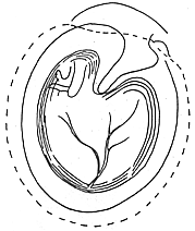
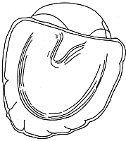
Figure 2: Tympanic membrane repair (external grafting)
(1) Scraping the epithelium around the perforation (dotted line) (2) Free skin graft or fascia covering the perforation externally
(2) Ossicular chain reconstruction Chronic otitis media often involves necrosis of the ossicles, most commonly the long process of the incus. During surgery, the ossicular chain should be reconstructed. If the long process of the incus is necrotic, the incus body can be lowered to connect with the stapes. If the incus is absent, the malleus long process can be repositioned to connect with the stapes, or an artificial incus can be used. If only the stapes or footplate remains, a columella or small tympanoplasty can be performed. In recent years, successful cases of allograft ossicles and total ossicular chain transplants have been reported, but sourcing materials remains challenging, limiting widespread adoption.
(3) Epitympanotomy with Antrum Approach: Performed under local or general anesthesia. An endaural incision is made, and the superior skin flap of the external auditory canal, along with the posterior portion of the tympanic membrane, is reflected anteriorly and inferiorly to expose the lateral wall of the epitympanum. The lateral wall is removed using a bone chisel or electric drill to open the entrance of the antrum, exposing all areas of bony destruction and cholesteatoma lesions. All necrotic mucosa and granulation tissue are cleared, along with any necrotic ossicles and cholesteatoma. The head of the malleus is excised. After irrigation and hemostasis, the skin flap of the external auditory canal is repositioned and pressed against the antrum area. Additionally, temporalis fascia or periosteum may be harvested and placed beneath the perforation of the tympanic membrane, followed by external packing with iodoform gauze. This procedure is also referred to as a modified radical mastoidectomy (Figure 3).
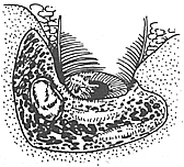
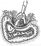
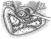
Figure 3 Removal of cholesteatoma in the epitympanum and mastoid antrum
(4) Tympanotomy The lesions are removed while preserving the posterior-superior wall of the external auditory canal and the tympanic cavity. The surgery is divided into three approaches: anterior approach, posterior approach, and combined approach.
① Anterior Approach This method is used when the lesion is limited to the epitympanum and the mastoid antrum is normal. An endaural incision is made, and the anterior and posterior skin flaps of the external auditory canal are reflected forward and downward to expose the epitympanic wall. The epitympanum is opened without entering the mastoid antrum, and only the lesions in the anterior-superior tympanum and the Eustachian tube orifice are removed, followed by tympanic membrane and ossiculoplasty.
② Posterior Approach A postauricular incision is made, and a simple mastoidectomy is performed. After clearing the lesions in the mastoid and mastoid antrum, under the microscope, an electric drill is used to extend forward and downward from the mastoid antrum, preserving the tympanic sulcus and the thin posterior bony wall of the external auditory canal. Between the short process of the incus and the chorda tympani nerve, a small drill bit is used to remove the posterior wall of the facial recess, exposing the posterior-superior region of the mesotympanum. The lesions in the facial recess and around the round window are cleared, followed by tympanic membrane and ossicular reconstruction as needed (Figure 4).
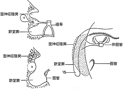
Figure 4 Relationship between the facial recess and the tympanic sinus
③ Combined Anterior-Posterior Approach Also known as the mastoidectomy with preservation of the posterior wall of the external auditory canal. In 1957, Jansen advocated this combined approach to preserve the posterior bony wall of the external auditory canal for the thorough removal of lesions and improvement of hearing in the treatment of type II or III chronic otomastoiditis. However, this requires advanced microsurgical skills in otology; otherwise, otitis media may not be cured. According to Jansen's report of 1000 surgeries, the recurrence rate of cholesteatoma was 2.2% (Figure 5).
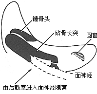
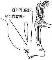
Figure 5 Jansen's Combined Anterior-Posterior Approach Surgery
(5) Radical Mastoidectomy Performed under general anesthesia, local anesthesia can also be attempted. If the mastoid is underdeveloped, an endaural incision is used; generally, a postauricular incision is employed. The mastoid is exposed, and the entire diseased mastoid air cells are removed using an electric drill or chisel. Granulation tissue and sebaceous cysts are thoroughly scraped away (Figure 6). For a radical procedure, the mastoid antrum should be enlarged, and the lateral bony wall (i.e., the bridge) should be removed. The posterior bony wall of the external auditory canal should be lowered to no less than the level of the incudal fossa; otherwise, the vertical segment of the facial nerve may be injured. The surgery should be performed under clear visualization to avoid injuring the tegmen plate, sigmoid sinus plate, facial nerve, or semicircular canals. If intracranial complications are suspected, even if the bony wall is intact, the bone plate should be drilled open for exploration. If the lesion is not severe and the skin of the external auditory canal is normal, the skin flap of the external auditory canal can be longitudinally split at the mastoid antrum, divided into upper and lower flaps, and pushed forward. Alternatively, it can be completely excised. The lesions in the tympanic cavity should then be cleared, preferably under microscopic visualization. Except for preserving the stapes and the round window niche, necrotic membranes, granulation tissue, necrotic ossicles, sebaceous cysts, and the tensor tympani muscle in the tympanic cavity should all be removed. If the tympanic lesions are mild, hearing loss is minimal, and the Eustachian tube functions normally, and if a tympanoplasty is anticipated at an intermediate stage [second stage], the removal of diseased tissue can be slightly conservative. Otherwise, all lesions should be thoroughly cleared, especially the membranous and granulation tissue at the tympanic orifice of the Eustachian tube. Incomplete removal is often the main cause of postoperative persistent discharge. Generally, after a radical mastoidectomy, the external auditory canal, tympanic cavity, mastoid antrum, and mastoid form a large cavity. The preserved posterior wall skin flap of the external auditory canal is divided into upper and lower flaps and folded into the mastoid cavity, fixed to the postauricular soft tissue. A Thiersch graft from the thigh is then placed in the mastoid-tympanic cavity, and the cavity is packed with iodoform gauze. The iodoform gauze is removed 9–10 days postoperatively, and 4% boric acid alcohol ear drops are applied. The ear usually dries within 1–2 weeks. The drawback of leaving a large cavity postoperatively is that exposure to hot or cold air or water may cause vertigo and headache. The grafted skin is very thin and poorly vascularized, making it prone to epithelial desquamation, ulcer formation, recurrent discharge, or epithelial accumulation leading to sebaceous cysts. To eliminate the postoperative cavity, mastoid cavity obliteration was widely practiced in the 1960s. This involved preserving the intact posterior wall skin of the external auditory canal and filling the mastoid cavity with a pedicled temporalis muscle flap or sternocleidomastoid muscle flap. Autologous rib or iliac bone grafts were also used, and even heterologous bone was employed. Long-term follow-up showed that although some bone grafts became infected and fell out or muscle flaps were absorbed, re-creating the cavity, and a few cases of recurrent sebaceous cysts occurred, most patients had no scab formation postoperatively, and infections were rare, making the technique still valuable.
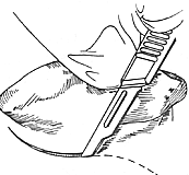
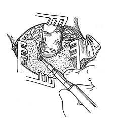
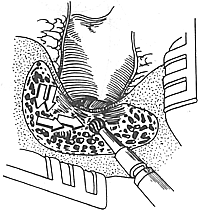
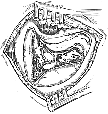
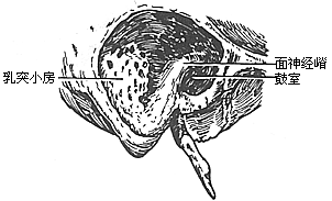
Figure 6 Simple Mastoidectomy and Radical Mastoidectomy
(1) Dotted line indicates the incision in children to avoid injury to the facial nerve. (2) The mastoid cortex and air cells are removed using a chisel or electric drill. (3) Arrows indicate the drilling direction to avoid injury to the semicircular canals, dural plate, and sigmoid sinus plate. (4) Cavity of simple mastoidectomy. (5) Cavity of radical mastoidectomy.
Common complications of radical mastoidectomy include: ① Traumatic deviation of the mouth, caused by unfamiliar anatomy, improper surgical technique, or congenital abnormalities of the facial nerve anatomy, which can result in partial or complete injury, with 80% occurring in the tympanic segment. ② Injury to the horizontal semicircular canal or removal of the stapes, leading to symptoms such as vertigo, nausea, and vomiting. If secondary infection occurs, it may result in permanent total deafness. ③ Surgical exposure of the tegmen plate or sigmoid sinus plate, causing intracranial infections such as meningitis. ④ Injury to the jugular bulb or internal carotid artery, resulting in massive hemorrhage, occasionally seen in patients with large cholesteatomas or severe osteomyelitis. ⑤ Incomplete opening of the bony bridge or insufficient lowering of the posterior bony wall of the external auditory canal, leading to granulation tissue proliferation and subsequent scar formation, stenosis, or even atresia. ⑥ The most common complication is cholesteatoma. If granulation tissue, osteitis, or other diseased tissues are not completely removed, persistent postoperative purulent discharge will occur.


