| disease | Oral Leukoplakia |
| alias | Leukoplakia, Oral Leukoplakia |
Leukoplakia refers to white or gray-white keratotic patch-like lesions that occur only on the mucous membrane. These patches on the oral mucosa cannot be wiped off and cannot be classified into other diseases clinically or histopathologically. It is a common non-contagious chronic disease. It can occur in any part of the oral mucosa but is most common on the cheeks and tongue. A 1980 national survey in China showed a prevalence rate of 10.47% (including smoker's patches). For a long time, all white patches occurring on the oral mucosa were referred to as "leukoplakia," leading to the conflation of many white lesions with leukoplakia, resulting in inappropriate epidemiological and treatment outcomes. In addition to white coloration, leukoplakia can also present as mixed red-and-white lesions. It should be clarified that leukoplakia is a clinical term based on macroscopic observation. The histopathological changes should meet the characteristics of precancerous lesions—epithelial dysplasia—rather than simple epithelial hyperplasia.
bubble_chart Etiology
Local irritants play a significant role in the pathogenesis of leukoplakia. Smoking is a common cause, with 80–90% of leukoplakia patients having a smoking habit, and the affected areas often coincide with the sites of tobacco irritation. Other factors such as chewing Areca Seed, alcohol, vinegar, spicy or hot foods, ill-fitting dental prostheses, residual crowns, and roots can also induce leukoplakia. Among systemic factors, Candida albicans infection, iron-deficiency anemia, deficiencies of vitamin B12 and folic acid, syphilis, as well as radiation and xerostomia, are closely associated with leukoplakia. The condition is more prevalent in middle-aged and elderly individuals, with a higher incidence in males than females (male:female = 13.5:1).
bubble_chart Pathological ChangesWhite lesions are precancerous damages, which microscopically show typical epithelial dysplasia, including nuclear hyperchromatism, increased mitosis, loss of polarity, altered nuclear-cytoplasmic ratio, and dyskeratosis. As for the surface morphology of the epithelium, such as wrinkled or verrucous appearances, although they are not indicative of precancerous changes, they should be highly vigilant.
bubble_chart Clinical Manifestations
1. Patch-like: White or gray-white homogeneous, hard patches appear on the oral mucosa, with a tight texture and varying shapes and sizes. Grade I lesions may be raised or uneven. Notably, the size of the lesion does not correlate with the likelihood of cancerous transformation; sometimes, even a rice-sized patch may already be cancerous. Macroscopically, patch-like lesions are often difficult to distinguish from Candida-associated leukoplakia, but the former feels harder upon palpation. (Color Figure 1)
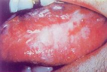
Color Figure 1: Leukoplakia
2. Granular: Also known as granular-nodular leukoplakia, it commonly occurs in the corner of the mouth. The lesion often forms a triangle with the base at the commissure. The color is mixed red and white, with the red areas representing atrophic erythroplakia, and the surface "speckled" with nodular or granular white patches. Hence, it has many synonyms: nodular-granular leukoplakia, granular erythroplakia, or non-homogeneous erythroplakia. Candida infection is frequently found in this type.
3. Verrucous: The lesion is raised with an uneven surface, accompanied by papillomatous or spiny projections, and feels slightly hard upon palpation (Color Figure 2). Except when located on the gums or palate, the base is not significantly hardened, but the affected area feels noticeably rough. Pain often occurs due to ulcer formation.
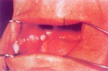
Color Figure 2: Verrucous Leukoplakia
4. Wrinkled: Mostly seen on the floor of the mouth and ventral tongue, also known as floor-of-mouth leukoplakia or sublingual keratosis. It can occur simultaneously in both areas or separately, even extending to the lingual gingiva. The lesion varies in size, with a surface resembling crumpled white paper and a soft base. Aside from a rough sensation, there are usually no obvious symptoms initially, and it is more common in women. Biopsy is necessary for diagnosis, as lesions in these areas often exhibit macroscopic "peaked projections" and similar microscopic features.
Any of the above types may be classified as "ulcerative" when ulceration occurs. Ulceration is a sign of further progression of precancerous lesions. Moreover, clinical diagnosis of each type must be confirmed by pathological examination to guide treatment selection.
The common sites of leukoplakia are the oral mucosa of the cheeks, dorsum and ventral surface of the tongue, lips, palate, floor of the mouth, and gums, but it can also occur in other areas. Certain types have relatively specific locations: granular leukoplakia is often found in the buccal mucosa of the commissural area; corrugated leukoplakia is commonly seen on the floor of the mouth and ventral surface of the tongue; and verrucous leukoplakia frequently appears on the gums. The affected areas of leukoplakia differ from those of leukokeratosis, and there are no similarities in morphology or texture. Candida-associated leukoplakia, in addition to microabscesses and epithelial dysplasia, can be identified by periodic acid-Schiff staining or culture methods to detect pathogens within the tissue. The distinction between leukoplakia and leukokeratosis can be made based on the site of occurrence, the texture of the lesion, and the clarity of the lesion's borders, with histological examination providing further confirmation.
bubble_chart Treatment Measures1. The primary measure is to remove irritants, such as quitting smoking, abstaining from alcohol, and avoiding hot or spicy foods. Residual roots, crowns, and poorly fitted restorations should also be removed. Harsh medications like phenols or silver nitrate must never be used to treat leukoplakia.
2. If the lesion persists after confirming the cessation of harmful habits like smoking, drug therapy may be considered, and further investigation into the disease cause should be conducted.
3. Oral administration of vitamin A (50,000 U daily) or retinoic acid (35–50 mg/d) may initially cause headache or dizziness, in which case the dose should be reduced. Adaptation usually occurs within a few days. From the second or third week onward, the dose can gradually increase to 30–60 mg daily, divided into three doses, with a treatment course of about 1–2 months. Common side effects include dry lips and alopecia areata. It is contraindicated in patients with coronary heart disease, liver or kidney dysfunction, or hyperlipidemia.
A 0.2% retinoic acid solution is suitable for topical application but should not be used on lesions with congestion or erosion. Before application, saliva should be wiped dry, and a fine brush should be used to apply a small amount of solution along the white area, avoiding the vermilion mucosa. Ointment formulations are unsuitable as they do not adhere well to the moist oral mucosa. Newer formulations like isotretinoin and arotinoid function similarly to retinoic acid, with the main advantages being a lower dose (0.5 mg/kg daily), reduced toxicity, and fewer side effects (dry lips, alopecia areata), though they have teratogenic effects.
4. Cod liver oil can be applied to leukoplakia long-term, 2–3 times daily, with a treatment course of 1–2 months, but excessive force should be avoided. Cod liver oil may also be taken orally, or vitamin A (50,000 U) can be administered. Close follow-up is necessary during conservative treatment.
5. For persistent leukoplakia that does not resolve with treatment, or if rhagades, ulcers, induration, or significant thickening are observed, or if precancerous changes are confirmed, surgical excision should be performed as early as possible.
1. White edema (leukoedema) is more common in middle-aged and older males than females, and is more likely to occur in individuals with full cheeks. White edema rarely causes subjective symptoms, so patients seldom seek medical attention for it. However, it is frequently observed in clinical dental practice. White edema may be a normal variation of the mucous membrane or could be related to stimuli such as smoking, alcohol, or hot food. The bilateral buccal mucosa appears semi-translucent and pale, resembling skin that has been soaked in water for too long. The interdental line area of the buccal mucosa is often the most prominent site of edema. The edematous area has a smooth surface with indistinct boundaries and may sometimes extend to the corners of the mouth and the upper and lower lips. Occasionally, noticeable edema may lead to the formation of several vertical or irregular folds. Palpation reveals a soft texture without tenderness. White edema generally does not require treatment.
Microscopic findings of white edema mainly include significant thickening of the spinous layer without a keratin layer, swelling of the spinous cells (swelling of eyelid), which becomes more pronounced toward the superficial layers, disappearance or condensation of nuclei, unstained cytoplasm, normal deep spinous and basal cells, irregular elongation of epithelial rete ridges, and slight inflammatory cell infiltration in the connective tissue.
2. White folded gingivostomatitis is present at birth but is not obvious. It begins to develop rapidly during adolescence and gradually stabilizes without worsening with age. Folded gingivostomatitis is a rare autosomal dominant genetic disorder that can affect not only the oral mucosa but also the nasal cavity, anus, and vulva. This condition is also known as white spongy nevus.
The lesions appear gray-white or milky-white, presenting as folded, spongy, scaly, thickened, and soft tissue. On palpation, these areas retain the flexibility and elasticity of the mucosa but feel spongy. Buccal mucosal lesions are more common, but other areas may also be affected, sometimes involving the entire oral mucosa. Smaller scaly tissues can be peeled off painlessly, revealing a light pink, smooth, non-bleeding "surface" resembling normal mucosa. Oral administration of retinoids shows significant therapeutic effects.
Microscopic findings of white folded gingivostomatitis include marked thickening of the squamous epithelium (up to 40–50 layers or more) without a granular layer, parakeratosis, swelling of the spinous cells (swelling of eyelid), which becomes more pronounced toward the surface, unstained cytoplasm, and slight inflammatory cell infiltration in the connective tissue.


