| disease | Bladder Injury |
The bladder is an organ that stores and excretes urine. It expands or empties depending on the amount of urine stored. In infants and children, the bladder is located above the pubic arch in the lower abdomen. In adult males, the bladder is situated between the pubic bone and the rectum. It connects inferiorly with the prostatic urethra and is posteriorly adjacent to the seminal vesicles and the ampulla of the vas deferens. Between the bladder and the rectum lies the rectovesical pouch. In females, the bladder is posterior to the uterus, with the vesicouterine pouch located between them. Therefore, the female bladder is positioned more anteriorly and lower than the male bladder, and the peritoneal reflection covering the posterior wall of the bladder is higher in females due to its connection with the uterus. The bladder wall below the urachus directly contacts the anterior abdominal wall without peritoneal covering. When the bladder is empty, only its upper edge is covered by the peritoneum, while the anterior, inferior, and lateral walls are not. When the bladder is full and distended, it rises into the lower abdomen, and the peritoneum covering the dome of the bladder also elevates. Thus, the position of the bladder and its relationship with surrounding organs can vary depending on age, gender, and the degree of urine filling. These anatomical and physiological characteristics of the bladder are closely related to the type, location, and extent of its injuries.
bubble_chart Etiology
Bladder injury mostly occurs when the bladder is filled with urine. At this time, the bladder wall is tense, the bladder area increases and rises above the pubic symphysis, becoming an abdominal organ, thus it is prone to injury. When the bladder is emptied, it is located deep in the pelvis, protected by surrounding fascia, muscles, pelvis, and other soft tissues, so except for penetrating injuries or pelvic fractures, it is rarely injured by external violence. According to the cause of injury, bladder injury can be divided into three categories:
(1) Closed injury: An overfilled or diseased bladder (such as tumors, ulcers, inflammation, diverticula) is prone to rupture due to external violence. This is commonly seen in cases of severe blows, kicks, falls, or accidental traffic accidents. When a pelvic fracture occurs, fracture fragments can also puncture the bladder. Drunkenness is one of the factors causing bladder rupture. When drunk, the bladder is often distended and filled, and the abdominal muscles are relaxed, making it prone to injury. Any disease that can cause urinary retention, such as urethral stricture, bladder stones or tumors, prostatic hypertrophy, and neurogenic bladder, can also be a predisposing factor for bladder rupture. When drunk or when the bladder already has lesions, bladder rupture can even occur without obvious external violence, which is called spontaneous rupture. Spontaneous bladder rupture is almost always intraperitoneal bladder rupture.(2) Open injury: Mainly seen in wartime, caused by firearms and sharp instruments, often combined with other organ injuries, such as rectal injury and pelvic injury. Generally speaking, bladder injuries caused by shrapnel or stab wounds entering from the buttocks, perineum, or thigh are mostly extraperitoneal, while those caused by penetrating trauma through the abdomen are mostly intraperitoneal.
(3) Surgical injury: Seen in cystoscopy, lithotripsy, intravesical B-ultrasound examination, transurethral prostatectomy, bladder neck electroresection, transurethral bladder cancer electroresection, childbirth, pelvic and vaginal surgery. It can even occur during inguinal hernia (bladder sliding hernia) repair. The main reason is improper operation, and bladder lesions themselves increase the chance of such injuries.
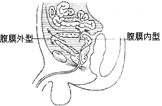
bubble_chart Pathological ChangesGrade I bladder contusion is limited to the wall layer of the bladder, with no urine extravasation, and does not cause serious consequences. Clinically, the main type of bladder injury encountered is rupture. Based on the relationship between the rupture location and the peritoneal membrane, it can be divided into two types: intraperitoneal rupture and extraperitoneal rupture (Figure 1).

Figure 1 Types of bladder rupture and the direction of urine extravasation
1. Extraperitoneal bladder rupture: The bladder wall is ruptured, but the peritoneal membrane remains intact. Urine extravasates into the surrounding tissues of the bladder and the retropubic space, extending to the subcutaneous tissue of the anterior abdominal wall, spreading along the pelvic fascia to the pelvic floor, or along the loose tissue around the ureter to the renal region. The injury site
is often located on the anterior wall of the bladder. Extraperitoneal bladder rupture is often associated with pelvic fractures. In one study of 1798 cases of pelvic fractures, 181 cases (10%) involved bladder rupture. Among another group of 259 cases of bladder rupture caused by pelvic fractures, 212 cases (82%) were extraperitoneal ruptures, and 47 cases (12%) were intraperitoneal ruptures.
2. Intraperitoneal bladder rupture: The bladder wall is ruptured along with the peritoneal membrane, and the bladder wall opening communicates with the abdominal cavity, allowing urine to flow into the abdominal cavity, causing peritonitis. The injury site is often located on the posterior wall and dome of the bladder.
In a group of 100 cases of bladder rupture, 50% were extraperitoneal, 30% were intraperitoneal, and 20% involved both types.
bubble_chart Clinical ManifestationsGrade I bladder wall contusion presents only with lower abdominal pain, a small amount of terminal hematuria, and resolves spontaneously within a short period. Symptoms are more pronounced in cases of full-thickness bladder rupture. The manifestations vary depending on the location and size of the rupture, the time elapsed since the injury, and whether other organs are also injured. Intraperitoneal and extraperitoneal ruptures each have their own specific signs. Generally, bladder rupture may present with the following symptoms:
(1) Shock: Severe trauma, pain, and significant blood loss are the main causes of shock. In cases of extensive trauma with injury to other organs, such as pelvic fracture, fragments from the fracture may puncture the lower abdominal and pelvic blood vessels, leading to severe blood loss and shock.
(2) Pain: Pain in the lower abdomen or pubic region, along with abdominal wall rigidity, is particularly noticeable when the pelvis is compressed in cases of pelvic fracture. Hematuria that extravasates around the bladder and into the retropubic space can cause local swelling. If secondary infection occurs, leading to cellulitis and sepsis, the symptoms become more severe. If urine leaks into the abdominal cavity, symptoms of peritonitis may appear, with the peritoneum reabsorbing creatinine and urea nitrogen, leading to elevated blood levels of these substances.
(3) Hematuria and Urinary Dysfunction: Patients may experience urgency or the sensation of needing to urinate, but are unable to pass urine or only pass a small amount of bloody urine. In cases of full-thickness bladder rupture, urine may not be expelled from the urethra due to sphincter spasm, urethral blockage by blood clots, or extravasation of urine around the bladder or into the abdominal cavity. Catheterization in such cases may yield only a small amount of bloody urine.
(4) Urinary Fistula: In cases of open bladder injury, urine may leak from the wound. If the injury communicates with the rectum or vagina, bloody urine may be discharged through the anus or vagina. After the formation of a bladder-rectal fistula, fragments of feces and gas may be expelled during urination. Recurrent episodes can lead to severe urinary tract infections and stone formation.
(5) Advanced Stage Symptoms: Urine may leak from the wound or be discharged through the anus or vagina via a bladder-rectal or bladder-vaginal fistula. The bladder may shrink, leading to symptoms of frequent and urgent urination. Recurrent urinary tract infections may also occur.
Based on medical history, signs, and other examination results, bladder injury can be diagnosed. However, if accompanied by injuries to other organs, the symptoms of bladder injury may be obscured. Therefore, in cases of trauma to the lower abdomen, buttocks, or perineum, or closed injury to the lower abdomen, if the patient experiences urgency to urinate but cannot or only passes a small amount of hematuria, bladder injury should be considered. The following examinations are helpful in confirming the presence of bladder rupture.
(1) If the bladder is found to be empty with only a small amount of bloody urine during catheterization, bladder rupture with possible urine extravasation should be considered. A certain amount of sterile saline can be injected and then withdrawn after a short period. If the amount of fluid withdrawn is less than the amount injected, bladder rupture with urine extravasation should be suspected.
(2) After catheterization, a contrast agent can be injected through the catheter for bladder imaging to determine if there is bladder rupture, urine extravasation, and the site of extravasation. Sometimes, it may even be found that the catheter has passed through the bladder rupture into the abdominal cavity, thus confirming the diagnosis.
(3) Excretory urography: If the patient's condition permits, excretory urography can be performed to display the structure and function of the urinary tract.
(4) Abdominal paracentesis: If ascites is present, abdominal paracentesis can be performed. If a large amount of bloody fluid is withdrawn, the urea nitrogen and creatinine levels can be measured. If these levels are higher than those in the blood, it may indicate extravasated urine.
Other examinations, such as a pelvic X-ray, can determine the presence of pelvic fractures or foreign bodies; an abdominal X-ray can detect the presence of free gas under the diaphragm. Elevated blood urea nitrogen and creatinine levels may be due to the reabsorption of intraperitoneal urine and do not necessarily reflect renal function. If there is diagnostic uncertainty and clinical symptoms suggest possible bladder rupture, exploratory surgery should be performed as soon as possible. Especially for patients with intraperitoneal rupture, emergency surgical treatment is required.
bubble_chart Treatment Measures
The early treatment of bladder rupture includes comprehensive therapy, prevention and treatment of shock, emergency surgery, and infection control. Advanced stage treatment mainly involves bladder fistula repair and general supportive care.
(1) Management of Shock The prevention and treatment of shock are the most critical first aid measures and necessary preparations before surgery, including blood transfusion, fluid infusion, and the use of stimulants to quickly bring the patient out of shock. This situation is particularly common when accompanied by pelvic fracture.
(2) Emergency Surgery The treatment method depends on the location of the injury, the presence of infection, and whether there are associated injuries. The main goals of surgery are drainage of urine, control of bleeding, repair of the bladder rupture, and thorough drainage of extravasated fluid. If other organs in the abdominal cavity are also injured, appropriate treatment should be given simultaneously.
Surgical Steps: A midline suprapubic incision is made, followed by cutting through the underlying fascia and separating and retracting the rectus abdominis to expose the prevesical space. Extraperitoneal and intraperitoneal bladder ruptures are managed as follows:
1. Extraperitoneal Bladder Rupture A large amount of blood and urine extravasation can be seen in the prevesical space. After suction, the anterior wall of the bladder is revealed. Fractured pubic bones need not be meticulously examined. If fractured fragments or foreign bodies puncture the inferior epigastric vessels or bladder, these fragments should be removed, and the bleeding vessels ligated to stop bleeding. If necessary, the anterior bladder wall is incised to explore the interior of the bladder, confirming the location and size of the rupture. After removing non-viable tissue, the inner mucosal layer of the rupture must be sutured with absorbable sutures. Care should be taken to avoid suturing the ureters. If the patient is critically ill and the rupture is near the bladder neck, making meticulous suturing difficult, do not force a repair. Perform a suprapubic cystostomy and thoroughly drain the prevesical space, and the rupture may heal on its own. After repairing the bladder rupture, a indwelling catheter is left in place for about a week before removal. If there is urine extravasation in the abdominal wall, lumbar region, ischiorectal fossa, perineum, scrotum, or even the thigh, thorough incision and drainage must be performed to prevent secondary infection.
2. Intraperitoneal Bladder Rupture The peritoneum is incised, and the fluid in the abdominal cavity is suctioned. The dome and posterior wall of the bladder are explored to identify the rupture. The anterior bladder wall can also be incised below the peritoneal reflection to observe the interior of the bladder. After repairing the rupture, if there is no injury to the intra-abdominal organs, the peritoneum is sutured. A high cystostomy is created on the anterior bladder wall, and the prevesical space is drained.
(3) Advanced Stage Treatment This mainly involves managing bladder fistulas, which should only be performed after the patient's general condition improves and local acute inflammation subsides. Long-term bladder fistulas can lead to severe infection and contracture of the bladder, and appropriate preventive measures should be taken. The main surgical steps include excision of the fistula and scar tissue around the fistula opening, suturing of the fistula, and creation of a high suprapubic cystostomy. Colostomy should be closed only after complete healing of the bladder-rectal fistula. Bladder-vaginal and bladder-uterine fistulas should be repaired, with an additional cystostomy created suprapubically, and the prevesical space drained.


