| disease | Triatrial Heart |
Triatrial heart is a rare congenital cardiovascular malformation where the left atrium is divided into two parts by an abnormal fibromuscular septum or membrane. The proximal side of the septum, located posterolaterally and superiorly, receives blood from the pulmonary veins and is termed the accessory atrium or accessory chamber, also known as the third cardiac chamber. The distal side of the septum, situated anteroinferiorly, is the true left atrium, which connects to the left atrial appendage, mitral valve, and left ventricle. Communication between these two parts occurs through an opening in the septum. This anomaly can exist in isolation but is often associated with other malformations, most commonly atrial septal defect or total anomalous pulmonary venous return.
bubble_chart Etiology
It is generally believed that during embryogenesis, the common venous trunk fails to fuse with the left atrium, while the pulmonary venous trunk enlarges to form part of the left atrium, failing to integrate with the primitive left atrium to create an accessory chamber. Additionally, abnormal development of the primary atrial septum and an abnormal membrane within the left atrium divides it into an accessory chamber and the true left atrium.
The hemodynamic changes depend on the size of the intra-atrial septal membrane orifice and associated anomalies. In isolated cor triatriatum sinistrum, the hemodynamics resemble mitral stenosis. Cases with a left septal membrane orifice diameter of only a few millimeters can cause pulmonary venous stasis, pulmonary congestion, pulmonary edema, and pulmonary hypertension. If complicated by partial anomalous pulmonary venous return or an atrial septal defect located between the right atrium and the accessory atrial chamber, a left-to-right shunt occurs. If the atrial septal defect is adjacent to the true atrial chamber, a right-to-left shunt results.
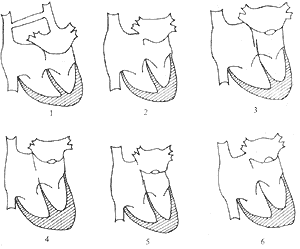
bubble_chart Clinical ManifestationsClinical Types: In 1964, Yoshitake Takeshita combined the classifications of Loeffler and Niwayama into three clinical types (Figure 1).

Figure 1 Schematic diagram of the anatomical classification of cor triatriatum
Type I: There is no communication between the accessory chamber and the true left atrium. The accessory chamber communicates via the foramen ovale or is accompanied by total anomalous pulmonary venous return, leading to early infant death.
Type II: There are one or several small channels between the accessory chamber and the true left atrium. From a clinical and surgical perspective, it is further divided into two subtypes: (1) No communication with the right atrium, with clinical manifestations resembling mitral stenosis. (2) Communication with the right atrium, with clinical manifestations resembling atrial septal defect or total anomalous pulmonary venous return.
Type III: There is a large communication between the accessory chamber and the true left atrium.
Clinical Manifestations: The timing of symptom onset is related to the size of the membrane orifice. In severe cases with a narrow orifice, grade III pulmonary congestion and tachypnea may appear shortly after birth, followed by severe pneumonia and congestive heart failure. In cases with a larger orifice, symptoms appear later, during infancy or childhood. Cases with a large orifice resemble atrial septal defects and may be clinically asymptomatic, with normal daily life and only mild dyspnea after exertion. In most cases, an ejection systolic murmur and diastolic murmur can be heard at the heart base, and sometimes a continuous murmur may be present due to a high pressure gradient between the proximal and distal sides of the severely obstructed orifice, with accentuated P2
bubble_chart Auxiliary Examination
X-ray examination: The heart is mildly to grade II enlarged, predominantly with right ventricular hypertrophy. The characteristic features include significant pulmonary hypertension without left atrial enlargement or only grade I enlargement, dilation of the superior vena cava, pulmonary interstitial edema, and prominence of the pulmonary stirred pulse segment indicating pulmonary arterial and venous hypertension.
Electrocardiogram: Right axis deviation and right ventricular hypertrophy are observed, with an elevated P wave suggesting right atrial hypertrophy.
Echocardiography: B-mode echocardiography reveals an abnormal septal membrane echo above the mitral valve within the left atrium. Pulsed Doppler ultrasound can demonstrate the abnormal septal membrane, visualize blood flow through the perforation in the membrane, and determine the size of the atrial septal defect, which is highly diagnostic.
Cardiac catheterization and angiography: A key feature is the elevated pulmonary stirred pulse wedge pressure measured by right heart catheterization, while the true left atrial pressure remains low or normal. In about one-third of cases, the catheter can pass through the atrial septal defect or foramen ovale into the left atrium after entering the right atrium. Left atrial angiography can reveal the presence of an abnormal septal membrane within the left atrium. If an accessory atrium is visualized, it remains non-contractile and maintains a constant morphology throughout the cardiac cycle. Routine cardiac catheterization and angiography are generally not necessary.
bubble_chart Treatment Measures
Once the diagnosis of cor triatriatum is confirmed, surgical treatment is the only method. It requires complete resection of the membrane under cardiopulmonary bypass, along with repair of the atrial septal defect and correction of other associated intracardiac anomalies. The surgical approach typically adopts the following two methods:1. Left atrial incision via the interatrial groove, suitable for infant cases without an atrial septal defect.
2. Right atrial incision with access to the left atrium through the atrial septum, suitable for children with a large atrial septal defect.
The surgical outcome is favorable, and pulmonary artery pressure may decrease to normal levels postoperatively. The mortality rate is high in infants and young children with congestive heart failure.
Congenital mitral stenosis: The hemodynamic changes are similar to those of cor triatriatum, making it difficult to differentiate clinically based on symptoms and signs. Echocardiography reveals left atrial enlargement without a septal membrane or intracardiac shunt, showing only the mitral stenosis lesion. Left atrial angiography demonstrates left atrial enlargement, delayed emptying, and the absence of a septal membrane or a third atrium.
Total anomalous pulmonary venous return: Chest X-ray shows a heart shadow resembling a "figure 8" or "snowman." Pulmonary angiography with contrast can reveal the abnormal pulmonary venous connections. Echocardiography displays the anomalous pulmonary venous return and atrial septal defect.
Left atrial myxoma: When the myxoma partially obstructs the mitral valve orifice, the clinical symptoms resemble those of mitral stenosis or cor triatriatum. Echocardiography shows an abnormal mass shadow in the left atrium that moves with the cardiac cycle.


