| disease | Gastric Mucosal Prolapse |
| alias | Prolapse of Gastric Mucosa |
Gastric mucosal prolapse is caused by the retrograde protrusion of abnormally relaxed gastric mucosa into the esophagus or its forward passage through the pyloric canal into the duodenal bulb, with the latter being more common clinically.
bubble_chart Etiology
The occurrence of this disease is mainly related to inflammation of the gastric antrum, and malignant cell infiltration of the gastric mucosa can also lead to this condition. When inflammation occurs in the gastric antrum, the submucosal connective tissue becomes looser, and the gastric mucosa and submucosa proliferate. If antral peristalsis increases, mucosal folds can easily be pushed into the pylorus, forming gastric mucosal prolapse. Any factors that cause intense gastric peristalsis, such as stress, tobacco, alcohol, or coffee bean stimulation, can serve as triggers for this disease. This condition often coexists with gastritis and duodenitis, and the relationship between them requires further study.
bubble_chart Pathological Changes
Since the vast majority of gastric mucosal prolapses are reducible, their presence may not be confirmed during surgery or autopsy. In cases of severe prolapse, the mucosal surface is congested and edematous, with possible erosion, ulceration, or polypoid hyperplasia, along with thickening of the pyloric region and widening of the pyloric orifice. Microscopically, the pyloric mucosa and submucosa exhibit congestion, edema, and glandular hyperplasia, accompanied by varying degrees of infiltration by lymphocytes, plasma cells, and eosinophils.
bubble_chart Clinical Manifestations
This condition is more common in men aged 30 to 60. Mild cases may be asymptomatic or only present with nonspecific symptoms such as abdominal distension and fullness, and belching. In cases where part of the gastric mucosa prolapses into the pylorus and cannot immediately reposition, symptoms may include dull pain, burning pain, or even colicky pain in the mid-upper abdomen, which may radiate to the back, often accompanied by nausea and vomiting. The onset of symptoms is often related to the patient's posture. For example, symptoms are more likely to occur when lying on the right side and less likely or even absent when lying on the left. Since eating can promote gastric peristalsis and facilitate gastric mucosal prolapse, symptoms often have a clear relationship with meals but lack obvious periodicity or rhythm. Alkaline medications may sometimes alleviate the pain, but their effect is far less significant than in peptic ulcers. Epigastric tenderness may be the only positive sign of this condition. When the prolapsed mucosa obstructs the pyloric canal, leading to incarceration or strangulation, a soft and tender mass may be palpable in the upper abdomen, accompanied by symptoms of pyloric obstruction, with or without gastrointestinal bleeding.
bubble_chart Auxiliary ExaminationSome patients test positive for fecal occult blood. During gastroscopy, the gastric antrum mucosa may appear normal, or show congestion and edema; sometimes, bleeding spots, erosions, or superficial ulcers may be observed. When the gastric antrum contracts, the gastric mucosa moves with peristalsis through the pylorus into the duodenum. Upon relaxation, the prolapsed gastric antrum mucosa can retract from below the pylorus back into the gastric cavity. X-ray gastrointestinal barium meal examination has definitive diagnostic value. When the patient is in the prone or right lateral position, a variable central filling defect can be seen at the base of the duodenal bulb. In typical cases, the pyloric canal appears widened, and the gastric mucosal folds pass through the pyloric canal into the duodenal bulb, causing the duodenal bulb to assume a "mushroom" or "parachute" shape (Figure 1).
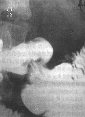
Figure 1: X-ray image of gastric mucosal prolapse showing the "mushroom" deformity at the base of the duodenal bulb.
This disease lacks characteristic clinical symptoms and signs, and endoscopic examination has limited diagnostic value. Definitive diagnosis mainly relies on X-ray barium meal examination. The condition also needs to be differentiated from peptic ulcer and chronic gastritis. The former presents with periodic, rhythmic abdominal pain unrelated to body position, and X-ray barium meal examination may reveal niche signs. The latter can be diagnosed with the aid of gastroscopy.
bubble_chart Treatment Measures
The disease is primarily managed with medical treatment, but there are no specific drugs available. General treatment includes eating small, frequent meals, quitting smoking and alcohol, and administering sedatives and anticholinergic drugs. Patients with pyloric obstruction or gastrointestinal bleeding should receive corresponding interventions. Surgical treatment is often considered when gastric mucosal prolapse causes pyloric incarceration or is complicated by massive gastrointestinal bleeding.

This disease should be differentiated from pedunculated gastric polyps prolapsing into the pyloric canal, hypertrophic pyloric stenosis, and gastric cancer.


