| disease | Cerebellar Tonsillar Herniation |
Cerebellar tonsillar herniation, also known as Arnold-Chiari malformation, is a developmental abnormality of the central nervous system often associated with basilar invagination.
bubble_chart Pathological Changes
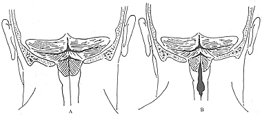
Cerebellar tonsillar herniation is caused by developmental abnormalities of the midline structures in the posterior cranial fossa during the embryonic period. The main pathological changes include the cerebellar tonsils elongating downward in a tongue-like shape, protruding through the foramen magnum of the occipital bone along with the lower segment of the medulla oblongata and entering the spinal canal. The adjacent pons and cerebellar vermis also shift downward, which may lead to deformation of the aqueduct and the fourth ventricle, as well as a series of changes such as narrowing of the subarachnoid space at the foramen magnum and the beginning of the spinal canal. In some cases, the tonsils may descend as low as the axis vertebra or even lower. In severe cases, part of the lower vermis may also herniate into the spinal canal. Due to these changes, cranial nerves such as the glossopharyngeal, vagus, accessory, and hypoglossal nerves, as well as the upper cervical spinal nerve roots, may be stretched and displaced downward. The foramen magnum and the upper cervical spinal canal become obstructed, leading to hydrocephalus. If this condition is accompanied by spinal meningocele or other malformations in the foramen magnum region, symptoms may appear earlier and be more severe than in isolated cases. Based on pathological changes, it can be classified into Type A (associated with syringomyelia) and Type B (simple tonsillar herniation) (Figure 1).

Figure 1 Arnold-Chiari malformation
A: Type A malformation; B: Type B malformation
bubble_chart Clinical Manifestations
Due to compression of the brainstem and upper cervical spinal cord, ischemia of neural tissue, involvement of cranial-spinal nerves, and obstruction of cerebrospinal fluid circulation, the following symptoms usually occur.
Symptoms of medulla oblongata and upper cervical spinal cord compression: Manifest as varying degrees of motor and sensory disturbances in one side or all four limbs, hyperactive tendon reflexes, positive pathological reflexes, dysfunction of the bladder and anal sphincter, difficulty breathing, etc.
Symptoms of cranial nerves and upper cervical nerves: Manifest as facial numbness, diplopia, tinnitus, hearing impairment, difficulty speaking and swallowing, pain in the lower occipital region, etc.
Cerebellar symptoms: Manifest as ocular tremor, unsteady or ataxic gait, etc.
Signs of intracranial hypertension: Due to compression and flattening of the brainstem and upper cervical segments, accompanied by adhesion and thickening of the surrounding arachnoid membrane, cysts may sometimes form. The medulla oblongata and cervical spinal cord may develop secondary syringomyelia or cervical hydromyelia due to ischemia caused by compression and the effects of cerebrospinal fluid pressure.
To clarify the diagnosis and differential diagnosis, magnetic resonance imaging (MRI), CT scanning, and vertebral angiography may be performed. For patients with increased intracranial pressure, sudden respiratory arrest should be monitored during the examination, so caution is required and emergency measures should be in place.
bubble_chart Treatment MeasuresThis disease does not always require surgical treatment upon diagnosis, as a significant number of cases present with mild clinical symptoms. Close observation is recommended for younger or older patients. Surgery should only be performed for those with severe symptoms and signs. The goals of surgery are to relieve compression on neural tissues, reconstruct cerebrospinal fluid circulation pathways, and stabilize unstable occipitocervical joints.
**Surgical Indications** (1) Compression of the medulla oblongata and upper cervical spinal cord; (2) Progressive worsening of cerebellar and cranial nerve symptoms; (3) Cerebrospinal fluid circulation障碍, increased intracranial pressure; (4) Dislocation of atlantoaxial vertebrae or instability.
**Surgical Methods** The primary procedures include partial resection of the occipital bone to enlarge the foramen magnum and decompression via resection of the posterior arch of the atlas. The dura mater should be widely incised, adhesions separated, and the median aperture of the fourth ventricle explored. If adhesions cause obstruction, careful separation and dilation should be performed to restore patency. If obstruction cannot be relieved, a shunt procedure to reconstruct the cerebrospinal fluid circulation pathway should be considered. For unstable dislocation of atlantoaxial vertebrae, occipital bone and cervical fusion should be performed.
The surgical procedure is described as follows:
**Surgical Steps**
Preoperative preparation, anesthesia, and positioning are the same as for posterior cervical spine surgery.
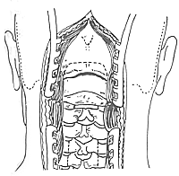
1. **Incision and Exposure**: The incision includes both the occipital and cervical regions, extending vertically from the midline of the occipital bone protuberance downward to the C6–7
level, exposing the occipital bone, posterior edge of the foramen magnum, posterior arch of the atlas, and the spinous processes of the second to sixth or seventh cervical vertebrae (Figure 1). The exposure method is as previously described.
**Figure 1** Exposure of the Occipitocervical Region
2. **Enlargement of the Foramen Magnum and Resection of the Posterior Arch of the Atlas**: The exposure and resection methods are as previously described. The extent of the foramen magnum incision should be sufficiently large. Typically, 2.5 cm to 3.0 cm of the occipital bone is removed upward from the foramen magnum, with a width of 3.0 cm to 4.0 cm, to ensure adequate decompression of the occipitocervical region.
3. **Syringosubarachnoid Shunt**: Based on magnetic resonance imaging, select the segment with prominent syringomyelia. Take care to protect C2 and C3 . The preferred sites are at C4–5 or C5–6 , or the C4, 5–6 segments. After laminectomy to expose the dura mater, incise the dura and retract the edges with fine sutures to expose the swollen spinal cord. Observe the distribution of surface vessels and select an avascular area. Insert a fine needle into the spinal cord and gently aspirate to confirm the presence of yellow fluid, indicating the location of the syrinx. Use a sharp blade to make a 2.0 cm to 3.0 cm incision at the needle site (avoid excessive force to prevent injury). Insert a silicone tube or strip (approximately 3.0 cm long and 1.0 mm in diameter) into the spinal cord incision, advancing 2.0 cm to 3.0 cm into the cavity. The other end is secured to the arachnoid mater with 3-0 sutures (Figure 2). Finally, close the dura mater.

**Figure 2** Enlargement of the Foramen Magnum, Resection of the Posterior Arch of the Atlas, Occipitocervical Fusion, and Syringosubarachnoid Shunt
4. **Occipitocervical Fusion**.
**Intraoperative Considerations and Complications**
1. The effectiveness of occipitocervical decompression must be ensured. In addition to ensuring the foramen magnum incision is sufficiently large, the stability of the occipital bone fusion site should also be considered.
2. The dorsal spinal cord incision must be performed in an avascular area and at the site of syringomyelia to prevent bleeding from interfering with the surgical procedure and to ensure the accuracy of the drainage site. Any rough manipulation of the spinal cord is a factor that can cause spinal cord injury. The use of a surgical microscope for the procedure enhances precision and minimizes injury.
Postoperative Management
This surgical procedure is technically complex, involves extensive exposure, prolonged duration, and significant manipulation of the spinal cord. Postoperatively, administer dexamethasone and diuretics such as furosemide for 5-7 days. Routine prophylactic antibiotics should be used. Remove sutures on the 10th postoperative day, followed by immobilization with a head-neck-thorax cast for 3 months.


