| disease | Bilateral Facet Dislocation of Cervical Vertebrae |
Bilateral facet joint dislocation of the cervical spine is a typical flexion injury, which can occur at any segment from C2 to T1, but is most commonly seen below C4.
bubble_chart Pathogenesis
It is commonly seen in falls from heights, where the head and neck strike the ground, or from direct blows by heavy objects, causing the occipitocervical region to be subjected to flexion forces. Whiplash injuries can also lead to dislocation. When a high-speed vehicle suddenly brakes, the head and neck violently flex due to inertia. When the head and neck are subjected to flexion forces, the pivot point of the cervical spine's functional unit is located slightly posterior to the center of the intervertebral disc. Due to the flat articular surfaces of the cervical facet joints and their 45° angle to the horizontal plane, sudden flexion forces cause the inferior articular processes of the upper cervical vertebrae to shift forward, tearing the joint capsule, and then tilt backward and upward. As the external force continues and the inertia of the head's weight acts, the displaced inferior articular processes slide further forward, and the entire upper vertebral body moves forward accordingly. After the force dissipates, due to the contraction of neck muscles, three conditions may arise: first, spontaneous reduction may occur, potentially leading to cervical instability or a semi-dislocation state later; second, the cervical vertebrae dislocation may become elastically fixed, with the superior and inferior articular processes mutually supporting each other, forming a "perching" state where they oppose each other tip-to-tip (Figure 1); third, the inferior articular processes of the upper vertebral body may override the superior articular processes of the lower vertebral body, forming a "locked" state where the facet joints are back-to-back (Figure 2).

Figure 1 Bilateral facet joint dislocation caused by flexion forces, showing the tip-to-tip opposition of the superior and inferior articular processes
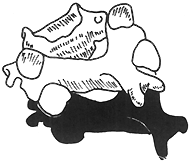
Figure 2 Bilateral facet joint "locked" state
bubble_chart Pathological Changes
The main pathological changes are the dislocation of the small articular processes on both sides of the injured segment. Due to hyperflexion trauma, all ligamentous structures of the motion unit at the injured segment, including the anterior longitudinal ligament, posterior longitudinal ligament, ligamentum flavum, interspinous ligament, and joint capsule, are torn. The intervertebral disc is no exception, with possible rupture of the annulus fibrosus and injury to the cartilage endplate. The superior vertebral body dislocates anteroinferiorly, which may be accompanied by fractures of the articular processes. The posterior structures, such as the nuchal ligament, are subjected to tensile stress and tear, leading to an increased interspinous distance in adjacent segments. Sometimes, it may also be accompanied by a grade I fracture of the vertebral body.
Due to the mutual displacement of the vertebral bodies, the morphology of the spinal canal is severely disrupted, resulting in a reduction of the canal at the corresponding level. The spinal cord is injured by the compression between the lamina of the superior vertebral segment and the posterior aspect of the inferior vertebral body. In severe cases, it can lead to a transverse injury of the spinal cord.
bubble_chart Clinical Manifestations
Local manifestation: The neck is in a forced position, with the head and neck forced into a forward flexion posture due to small joint locking and elastically fixed. Severe pain in the head and neck region is primarily caused by the significant increase in tensile and stretching stresses on the periarticular soft tissues during the dislocation state, exacerbating the pain. Due to the pain and mechanical abnormalities in the injured segment, neck muscles exhibit obvious spasms; the head cannot be passively moved; and there is widespread tenderness in the neck.
Neurological and spinal cord injury symptoms: Manifest as symptoms corresponding to the affected segment, such as quadriplegia, paraplegia, or incomplete paralysis. In cases with nerve root injury, symptoms include hypersensitivity, pain, or hypoesthesia in the skin area innervated by the affected nerve root.

bubble_chart Auxiliary Examination
The typical signs on lateral radiographs are: the dislocated vertebral body is displaced anteriorly by 2/5 of its anteroposterior diameter, the inferior articular process of the upper cervical vertebra is located at the top or anterior to the superior articular process of the lower cervical vertebra, and the distance between the two spinous processes is increased (Figure 1). On anteroposterior views, the uncovertebral joint relationship is disordered, and the interrelationships of the facet joints are unclear. Oblique views show deformation of the neural foramen. Tomography is more conducive to diagnosis.
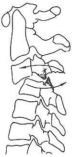
Figure 1 Radiographic features of bilateral facet dislocation
MRI examination can reveal deformation of the spinal canal and varying degrees of spinal cord compression. If there is injury or edema, it can also be manifested by changes in signal intensity.
In the history of trauma, it is important to understand whether there was any force causing extreme flexion of the cervical spine, such as a head-first fall injury, sudden braking while riding in a vehicle, or head and neck impact injuries in rugby players. Additionally, attention should be paid to whether there was any rotation of the head and neck at the moment of injury. In imaging examinations, characteristic X-ray findings are key to diagnosis.
First Aid Keep the airway unobstructed. If respiratory dysfunction occurs, immediately perform a tracheotomy or use a ventilator to maintain airway patency, sustain respiration, and administer oxygen appropriately.
Traction Reduction Skull traction should be utilized whenever possible. According to the mechanism of dislocation, continuous traction should first be applied in a slightly flexed position, and bedside fluoroscopy or X-rays should be used to confirm whether the facet joint lock has been resolved. Once the dislocation is corrected, the traction should be immediately adjusted to an extended position, maintaining a weight of 1.5–2 kg for 3–4 weeks, followed by fixation with a head-neck-thorax cast for 3 months. The goal of traction is reduction, and the following aspects must be noted during the reduction phase:
1. **Direction of Traction**: Avoid extension initially; start with slight flexion or a neutral position to prevent or exacerbate spinal cord injury.
2. **Method of Traction**: Avoid using a chin halter or manual traction; skull traction is safer and more effective.
3. **Traction Weight**: Begin with 3–4 kg and gradually increase. Take lateral cervical spine X-rays every 30 minutes to monitor reduction progress. The weight may be increased by 0.5 kg every half hour, but the total weight should not exceed 15 kg. Monitor blood pressure and pulse closely during reduction.
4. **Duration of Traction**: Do not rush the process. Traction reduction typically takes 5–8 hours; proceeding too quickly may cause iatrogenic injury.
**Surgical Reduction** Most bilateral cervical facet joint dislocations can be corrected via traction reduction. Surgical reduction is indicated in the following cases: - Failure to achieve reduction after 5–8 hours of traction, especially in cases over one week post-injury. - Progressive worsening of spinal cord injury symptoms during traction. - Old fracture-dislocation with incomplete paraplegia.
Surgical approaches include anterior and posterior methods.
**Posterior Approach**: Performed under skull traction with endotracheal intubation anesthesia. The patient is placed in a prone position with the head slightly flexed on a headrest. A midline posterior incision is made to expose and reduce the dislocation. If reduction is difficult, partially resect the superior articular process of the dislocated facet joint. Insert a blunt periosteal elevator into the inferior joint space and gently lever it into place under traction. If facet joint locking hinders reduction, resect the obstructing portion. If vertebral lamina or articular fractures encroach on the spinal canal, decompression via resection is necessary. For concurrent spinal cord injury, perform laminectomy decompression post-reduction. After reduction, extend the cervical spine and secure it with wire fixation.
**Anterior Approach**: Also performed under skull traction. The patient is supine, and access is gained via the medial border of the sternocleidomastoid muscle and the visceral space. After exposing the injured segment and precise localization, resect the damaged intervertebral disc. Under continuous skull traction, insert a periosteal elevator into the disc space, using the lower vertebra as a fulcrum. Gradually increase leverage while manually pressing the dislocated vertebra to reduce it. Post-reduction, carefully remove any bone fragments protruding into the spinal canal with a curette. Fill the decompressed space with autologous iliac bone graft for fusion. For added stability, an anterior titanium plate may be used. Due to the inherent uncertainty of anterior reduction, experience is crucial. Fluoroscopic guidance is recommended if available.
Postoperatively, administer intravenous dexamethasone and dehydrating agents to mitigate spinal cord irritation from surgical manipulation. Place sandbags beside the head to limit excessive neck movement. After suture removal, immobilize with a head-neck-thorax cast for 3 months, removing it only after radiographic confirmation of bone healing.
Prognosis is generally favorable unless spinal cord injury is present. For traumatic arthritis of the facet joints, interarticular fusion may be performed.


