| disease | Open and Close |
| alias | Open Bite |
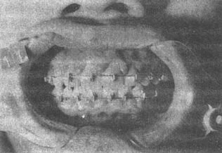
Open bite is relatively rare in clinical practice and is more commonly seen in the permanent dentition. It refers to a malocclusion where there is no vertical contact between the upper and lower teeth in both centric occlusion and non-centric occlusion positions.
bubble_chart Etiology
1. Genetic factors Heredity may contribute to open bite malocclusion, but there are differing opinions and ongoing debates on this issue, requiring further in-depth research.
2. Rickets Severe rickets is one of the significant causes of open bite malocclusion, leading to a large-scale wedge-shaped open bite with a wider anterior gap and a narrower posterior gap.
3. Oral habits Bad oral habits can disrupt the dynamic balance of the muscles in the oral and maxillofacial regions, resulting in open bite malocclusion. Such habits account for approximately 68.7% of the total disease causes of open bite.
The habit of protruding tongue is the most common cause of anterior open bite. Due to the thicker middle and thinner sides of the tongue, a spindle-shaped open bite gap often occurs, accompanied by mandibular protrusion and scattered anterior tooth spacing.
Additionally, habits such as thumb-sucking, object-biting, and lip-biting can lead to localized small open bites in different areas of the dentition.
4. Mandibular third molars In cases of mesially impacted or horizontally impacted mandibular third molars, they may occasionally push against the second molars, causing them to elongate and protrude beyond the occlusal plane, separating the other teeth. If combined with tongue-related factors, this can result in a large-scale open bite malocclusion.
bubble_chart PathogenesisDivided into three types:
1. The height of the anterior alveolar or jaw body is normal, while the height of the posterior alveolar or jaw body is excessive.
2. The height of the posterior alveolar or jaw body is normal, while the height of the anterior alveolar or jaw body is insufficient.
3. The height of the anterior alveolar or jaw body is insufficient, while the height of the posterior alveolar or jaw body is excessive.
Open bite is divided into 3 degrees: It refers to the vertical distance between the incisal edges of the upper and lower incisors, measured by the vertical distance from the incisal edge of the maxillary incisors to the incisal edge of the mandibular incisors.
Degree I: The vertical separation between the upper and lower open bite teeth is within 3mm.
Degree II: The vertical separation between the upper and lower open bite teeth is 3–5mm.
Degree III: The vertical separation between the upper and lower open bite teeth exceeds 5mm.
The range of open bite varies in size. Some cases involve only anterior open bite, some involve only localized posterior open bite, and severe cases may exhibit open bite where only the last pair of molars have occlusal contact in the entire dentition.
Open bite not only impairs cutting and chewing functions but also affects swallowing, speech, respiration, and facial appearance. The greater the degree and range of open bite, the more severe the impact on functions such as chewing.
In patients with open bite malocclusion, the height of the facial bones increases with the degree of open bite. Severe open bite significantly increases the lower third of the facial height, with an obtuse mandibular angle and often incomplete lip closure, frequently leading to periodontal and upper respiratory infections, thereby affecting overall health.
1. Different treatment designs are implemented based on the varying formation mechanisms.
(1) For cases with normal anterior alveolar height and excessive posterior alveolar height, if the patient is young, a posterior bite block can be worn to simply depress the posterior teeth. If necessary, a headgear and chin cap can be used in combination for vertical traction to stimulate the growth of the mandibular condyle.
(2) For cases with normal posterior alveolar height and insufficient anterior alveolar height, a light-wire or edgewise fixed appliance can be used. Bands are made on the four first permanent molars for anchorage, brackets are bonded to the labial surfaces of the teeth involved in the open bite, and the teeth are ligated together with archwires. Vertical elastics are used between the upper and lower anterior teeth to increase the anterior alveolar height (Figure 1).

A. Before treatment
B. After treatment
Figure 1 Vertical traction for anterior open bite correction
(3) For cases with insufficient anterior alveolar height and excessive posterior alveolar height, a fixed appliance can also be used to increase the anterior alveolar height and depress the posterior alveolar height.
2. For severe open bite in older patients with significant skeletal deformities, non-mechanical treatments may not be effective. A combination of orthodontic treatment and surgical intervention is recommended. Depending on the case, procedures such as maxillary anterior or major segmental osteotomy, mandibular body osteotomy, or mandibular ramus osteotomy may be selected.
1. Eliminate bad habits such as protruding tongue, licking teeth, tongue thrusting, thumb sucking, and lip biting. During the deciduous dentition or the initial stage of mixed dentition [first stage], if bad habits are eliminated before the age of 10, the  malocclusion may self-correct. Additionally, educate and persuade children to actively cooperate, and if necessary, use a habit-breaking appliance.

3. Extract the mandibular third molars that cause the malocclusion, allowing the overerupted mandibular second molars to self-reposition, and combine this with functional training of the masseter, temporalis, and medial pterygoid muscles.


