| disease | Uterine Rupture |
| alias | Rupture of Uterus |
Rupture of the uterus refers to the tearing of the uterine body or lower uterine segment during pregnancy or childbirth. It most commonly occurs during childbirth and is associated with factors such as obstructed labor, improper or difficult delivery procedures, misuse of uterine contraction agents, trauma to the pregnant uterus, and poor healing of uterine surgical scars. In rare cases, it may occur during advanced stages of pregnancy. Uterine rupture is one of the most severe obstetric complications, often leading to maternal and fetal mortality. Its incidence rate serves as an indicator of the quality of obstetric care in a region. In recent years, with the improvement in the number and expertise of obstetric professionals in China, as well as the establishment and gradual strengthening of the three-tier maternal and child healthcare network in urban and rural areas, the incidence of uterine rupture has significantly declined.
bubble_chart Clinical Manifestations
Uterine rupture can occur during the advanced stage of pregnancy before labor, but most cases happen during labor when childbirth encounters difficulties, manifesting as prolonged labor, the fetal head or presenting part failing to descend into the pelvis or being obstructed at or above the ischial spine level. Uterine rupture can generally be divided into two stages: threatened uterine rupture and uterine rupture.
1. **Threatened Uterine Rupture** During labor, when the descent of the fetal presenting part is obstructed, strong contractions cause the lower uterine segment to gradually thin while the uterine body thickens and shortens, forming a distinct annular depression between the two. This depression gradually rises to the level of the umbilicus or above, known as the **pathologic retraction ring**. At this stage, the lower segment becomes distended, with marked tenderness, and the round ligaments of the uterus are extremely tense, easily palpable, and tender. The mother complains of severe, unbearable lower abdominal pain, dysphoria, restlessness, crying out, and increased pulse and respiratory rates. Due to the compression of the bladder by the presenting part, urinary retention and hematuria may occur. The excessive frequency of uterine contractions impedes fetal blood supply, leading to changes in fetal heart rate or difficulty in auscultation. If this condition is not immediately relieved, the uterus will soon rupture at or below the pathologic retraction ring. (Figure 1)
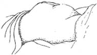
**Figure 1** Abdominal appearance in threatened uterine rupture
2. **Uterine Rupture** Based on the extent of rupture, it can be classified into **complete uterine rupture** and **incomplete uterine rupture**.
**(1) Complete Uterine Rupture** This refers to a full-thickness rupture of the uterine wall, connecting the uterine cavity with the abdominal cavity. At the moment of complete rupture, the mother often experiences a tearing, intense abdominal pain, followed by the cessation of uterine contractions and temporary pain relief. However, as blood, amniotic fluid, and the fetus enter the abdominal cavity, she soon feels generalized abdominal pain, with a rapid and weak pulse, tachypnea, and hypotension. Examination reveals generalized abdominal tenderness and rebound tenderness. The fetal body can be clearly palpated beneath the abdominal wall, while the uterus shrinks and lies beside the fetus. Fetal heart sounds disappear, and there may be varying amounts of fresh vaginal bleeding. The presenting part, which was previously descending or crowning, disappears (as the fetus enters the abdominal cavity), and the previously dilated cervix may retract. If the rupture occurs in the anterior uterine wall, the tear may extend forward, causing bladder rupture. Once uterine rupture is confirmed, vaginal examination to identify the rupture site is unnecessary. If rupture is caused by oxytocin injection, the mother may feel intense uterine contractions after administration, followed by sudden severe pain, the presenting part rising and disappearing, and abdominal findings as described above.
**Scar rupture of the uterus** may occur in the late stage of pregnancy (third trimester) but is more common during childbirth. Initially, there is mild abdominal pain and tenderness at the site of the uterine scar, indicating possible scar dehiscence, though the fetal membranes remain intact and fetal heart sounds are normal. Without immediate cesarean delivery, the fetus may enter the abdominal cavity through the rupture, leading to symptoms and signs similar to those described above.
**(2) Incomplete Uterine Rupture** This refers to partial or full-thickness rupture of the uterine muscle layer without perforation of the serosal layer, meaning the uterine cavity and abdominal cavity remain separate, and the fetus and its appendages remain within the uterus. On abdominal examination, tenderness is noted at the site of incomplete rupture. If the rupture occurs in the lateral uterine wall between the two layers of the broad ligament, a hematoma may form within the broad ligament, presenting as a gradually enlarging, tender mass on one side of the uterus. Fetal heart sounds are often irregular.The diagnosis of complete uterine rupture is generally not difficult and can be made based on medical history, childbirth process, clinical manifestations, and signs. Incomplete uterine rupture can only be detected under close observation. In some cases of advanced-stage pregnancy rupture, a definitive diagnosis can only be made when symptoms and signs of uterine rupture appear.
In certain difficult delivery cases, repeated vaginal examinations may lead to infection and peritonitis, presenting symptoms similar to those of uterine rupture. During vaginal examination, if the fetal presenting part remains high and the lower uterine segment is thin, bimanual palpation may give the impression that only the abdominal wall separates the fingers, sometimes leading to a misdiagnosis of uterine rupture. In such cases, the fetal body does not enter the abdominal cavity, and the pregnant uterus does not shrink but remains adjacent to the fetal body.
bubble_chart Treatment Measures
Signs of impending uterine rupture must be promptly addressed with effective measures to suppress uterine contractions, such as administering general anesthesia with ether or intramuscular injection of 100mg pethidine, to slow the progression of uterine rupture. A cesarean section should be performed as soon as possible, with careful examination during the procedure to determine whether uterine rupture has already occurred.
In cases where uterine rupture occurs before fetal delivery, even if the fetus is dead, vaginal delivery should not be attempted first, as this may enlarge the rupture, increase bleeding, and spread infection. Instead, a laparotomy should be performed promptly to remove the dead fetus. The decision on further management should consider the patient's condition, the location and extent of the rupture, the degree of infection, and whether the patient has children. If the uterine rupture is easily sutured, infection is not severe, and the patient's condition is poor, the rupture may be repaired. For patients who already have children, tubal ligation may be performed, while those without children may retain their fertility. Otherwise, a total or subtotal hysterectomy may be performed. For lower uterine segment ruptures, careful examination of the bladder, ureters, cervix, and vagina is necessary, and any injuries should be repaired immediately.Uterine rupture is often accompanied by severe bleeding and infection. Before surgery, blood transfusions, fluid resuscitation, and sodium lactate infusion should be administered, along with aggressive anti-shock treatment. During and after surgery, broad-spectrum antibiotics in larger doses should be used to control infection.
Improve the three-tier healthcare network, promote maternal health knowledge, strengthen prenatal check-ups, closely monitor the labor process, and prevent the occurrence of neglected difficult delivery. Those with a history of cesarean section or uterine incision surgery should be hospitalized in advance for delivery. The method of childbirth should be determined based on indications and previous surgical conditions. If there is no obstructive difficult delivery, vaginal childbirth can also be performed under close observation. The indications and methods for using uterine contractions such as oxytocin and prostaglandins should be strictly controlled to avoid misuse.


