| disease | Congenital Coxa Vara |
| alias | Developmental Coxa Vara |
Congenital coxa vara in children, also known as developmental coxa vara, occurs during early childhood. The neck-shaft angle of the femoral neck progressively decreases, presenting as a gradually worsening limp, which is one of the common causes of limping in children. Unilateral cases are more frequent than bilateral ones, with no significant differences in gender or race.
bubble_chart Etiology
The disease cause of congenital coxa vara is unknown, with a family history of inheritance and abnormal development of the epiphyseal plate. The calcification process on the medial side of the femoral neck is obstructed, leading to abnormal development of the medial femoral neck. Whether the cause is due to local vascular malformation or trauma remains undetermined.
bubble_chart Pathological Changes
The neck-shaft angle of the femoral neck is formed by the axes of the femoral neck and the femoral shaft. In children, the neck-shaft angle typically ranges from 135° to 145°, gradually decreasing to 120°–140° in adults. If the neck-shaft angle is less than 120°, it is referred to as coxa vara. In congenital coxa vara, a triangular bone defect or hypoplastic bone area is observed at the junction of the medial femoral head and the femoral neck. The apex of the triangular bone fragment connects with an osteoporotic band traversing the femoral neck, and pathological examination reveals delayed ossification of cartilage tissue. This region lies along the primary weight-bearing axis of the femoral neck, thereby reducing its load-bearing capacity, while the epiphyseal line is located proximal to this axis. With increasing age and weight, the child's standing and walking exert additional stress on the femoral neck, exacerbating its curvature and causing inward tilting of the femoral head epiphysis. This results in unfavorable shear and bending stresses on the osteoporotic cartilage tissue of the femoral neck, which intensify as the femoral neck bends. Severe coxa vara leads to progressive reduction of the neck-shaft angle, sometimes even approaching an acute angle. The osteoporotic band of the femoral neck widens, and the greater trochanter shifts upward until it contacts the ilium. Ultimately, the coxa vara deformity assumes a cane-like appearance.
bubble_chart Clinical ManifestationsChildren are generally asymptomatic before starting to walk, but develop a limp due to gluteus medius muscle laxity after walking. In cases of bilateral involvement, the gait resembles a duck's waddle, with the greater trochanter protruding outward and upward, and the femoral neck bending inward, resulting in limb shortening. Due to laxity of the gluteus medius and minimus muscles, the Trendelenburg sign is positive, and hip abduction, adduction, and rotation are limited. This condition differs from coxa vara caused by secondary epiphyseal slippage in childhood, ischemic necrosis of the femoral head, femoral cervical osteomyelitis, multiple osteochondrodysplasia, femoral neck fracture, and other diseases, but all present with a limping gait and should be differentiated from congenital hip dislocation. In this condition, the femoral head remains within the acetabulum, and the telescope test is negative. However, in the late stage (third stage), hip joint function is significantly restricted, and the incidence is relatively low (Figure 4).
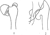
Figure 4 Congenital Coxa Vara Deformity
1. A radiolucent zone is present near the epiphyseal plate, forming a triangular bone fragment between the epiphyseal plate and the metaphysis.
2. The epiphysis fuses, resulting in a cane-shaped deformity.
Radiographic findings include: a decreased neck-shaft angle, a triangular bone fragment at the medial junction of the femoral neck and femoral head. This triangular fragment shows reduced density, appearing as an inverted V-shape, representing a dysplastic bone area with clear boundaries from surrounding bone. The medial boundary is the epiphyseal line of the femoral head, and the lateral boundary is a radiolucent zone of abnormal development. As age and weight increase, the weakened radiolucent zone widens and straightens, worsening the coxa vara. In advanced cases, the femoral head becomes distorted and elliptical, the acetabulum shallow, and the neck-shaft angle may decrease below 90°. The HE angle measurement (Figure 5) is normally around 25°; in coxa vara, it exceeds 25°. Serial measurements of the HE angle can assess the progression of coxa vara, determine the need for surgical correction, and estimate the degree of correction required to prevent recurrence of the deformity.
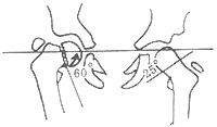
Figure 5 The HE angle is the angle between the YY′ line (Hilgenreiner line) connecting the Y-shaped cartilage of both acetabula and the epiphyseal plate line of the proximal femoral metaphysis (Hilgenreiner epiphyseal angle).
bubble_chart Treatment Measures
Congenital coxa vara patients experience non-physiological shear stress and bending stress between the femoral head and the femoral neck shaft. The treatment principle is to reduce bending stress during childhood growth to achieve normal or near-normal conditions, converting the shear stress between the femoral head and neck into physiological compressive stress.
For grade I coxa vara, non-surgical treatment may be adopted, while when the neck-shaft angle is less than 100°, surgical correction is often required to increase the neck-shaft angle, restore normal physiological compressive stress, and eliminate shear stress. The surgery involves subtrochanteric valgus osteotomy, transforming the originally vertical epiphyseal line into a horizontal one. Due to differences in osteotomy methods and fixation techniques, there are various surgical approaches. Below are the main ones introduced:
(1) Subtrochanteric Oblique Osteotomy: After anesthesia, the patient is placed in a supine position with the affected hip elevated. A longitudinal incision is made on the lateral side of the upper thigh to expose the greater trochanter and the upper third of the femur. An oblique osteotomy is performed slightly below the greater trochanter's epiphysis, extending downward to the lesser trochanter, forming an angle of approximately 35–45° with the femoral shaft. A bone groove is chiseled into the cancellous bone of the proximal osteotomy surface. The thigh is abducted, and the oblique tip of the distal femoral osteotomy is inserted into the groove of the proximal femoral trochanter. If insertion is difficult, the cortical bone edges of the distal femoral osteotomy's斜面 can be sharpened further, allowing the refined tip to fully insert into the groove. The degree of femoral shaft abduction depends on the preoperative coxa vara severity, and two screws are used to fix the upper femur and the medial cortical bone of the lesser trochanter. Postoperatively, skin traction is applied for 6–8 weeks before removal, followed by bed activity. Weight-bearing walking begins after X-ray confirmation of healing (Figure 1).
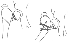
Figure 1 Subtrochanteric Oblique Osteotomy
(2) Subtrochanteric Wedge Valgus Osteotomy: The positioning and surgical approach are the same as above. A wedge-shaped bone block is removed from the subtrochanteric femoral shaft. The wedge angle is preoperatively calculated as the sum of the coxa vara angle and the wedge angle, equaling or slightly exceeding the normal neck-shaft angle. After wedge removal, the affected limb is abducted, and the osteotomy surfaces are aligned. A four-hole plate is bent to match the angle between the greater trochanter and femoral shaft and fixed laterally with screws. Postoperatively, a hip spica cast is applied until bone healing, after which the cast is removed, and walking begins (Figure 2).
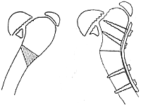
Figure 2 Subtrochanteric Wedge Valgus Osteotomy
(3) Inverted "V" Intertrochanteric Angle-Correcting Osteotomy: The patient is supine with the affected hip elevated. The adductor muscles are severed, and inverted V-shaped holes are drilled laterally at the greater trochanter, while transverse holes are drilled medially below the lesser trochanter. An osteotome connects the holes to complete the osteotomy. A curette removes cancellous bone from the proximal trochanter. The pelvis is stabilized, and the limb is fully abducted to embed the distal inverted V-shaped bone tip into the pre-chiseled proximal trochanteric groove. A half-hip spica cast is applied. Due to adductor muscle release and full abduction, postoperative recurrence is rare (Figure 3).
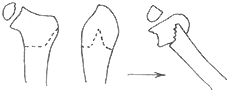
Figure 3 Inverted V Angle-Correcting Osteotomy
The following points should be noted during abduction osteotomy of the femoral trochanteric region. ① For children with coxa vara still greater than 100°, close follow-up is required. If the coxa vara progresses, early surgical treatment should be performed, as the later the surgery, the worse the functional recovery. ② After osteotomy, the hip should be fully abducted. Since coxa vara is mainly caused by abnormal development of the epiphyseal plate and is mostly progressive, if the adverse mechanical factors cannot be eliminated by surgical correction, there will still be varying degrees of recurrence postoperatively. Therefore, overcorrection should be performed during surgery to prevent recurrence of coxa vara. ③ During surgery, injury to the proximal femoral epiphysis should be avoided, otherwise it may lead to premature epiphyseal fusion. Bone grafting should not be performed in the lesion area of the femoral neck, as it not only fails to promote ossification but may also worsen the deformity.


