
| disease | Chronic Ethmoid Sinusitis |
Among the paranasal sinuses, the ethmoid sinus has the most complex anatomy, particularly the semilunar hiatus at the opening of the anterior ethmoid sinus and nearby structures such as the uncinate process and ethmoid bulla. These small protrusions and groove-like spaces in the middle meatus are collectively called the ostiomeatal complex. This area is where inhaled air impacts the nasal cavity and is also the most vulnerable site for bacteria, viruses, and inhaled allergens to invade. Whether due to infection or allergic reactions, the initial manifestation is swelling of the mucous membrane in this region, cessation of ciliary movement, and obstruction of ethmoid sinus ventilation and drainage, which then spreads to other sinuses. Due to poor drainage of the ethmoid sinus, inflammation is difficult to resolve and often becomes chronic.
bubble_chart Pathological Changes
Mucosal membrane lesions include three types: polyp, hypertrophy, and atrophy. Bone wall lesions can be classified into the following three types:
1. Proliferative bone lesions Due to congestion stimulation in the submucosal layer, proliferative osteitis occurs in the bone wall, causing the ethmoid bone wall to harden.
2. Atrophic bone lesions Long-term compression from polyps and hypertrophic mucosa on the ethmoid bone wall leads to insufficient blood supply, resulting in thinning or disappearance of the bone wall.
3. Ulcerative bone lesions Thrombophlebitis in the mucosal membrane spreads to the bone wall, causing necrosis of the ethmoid bone wall. The ethmoid cells may fuse into a large cavity filled with pus. If the infection is severe, intraorbital or intracranial complications may occur.
bubble_chart Clinical Manifestations
Chronic ethmoid sinusitis rarely occurs alone and its symptoms are atypical, with neuralgia, mental depression, and lack of concentration being more common. When the sinus opening is blocked, there may be a feeling of fullness at the root of the nose or around the eyes, along with a stuffy nose, olfactory dysfunction, and postnasal drip.
Clinical examination reveals polyps obstructing the middle nasal meatus, hypertrophy of the middle turbinate and the nodular part of the nasal septum, and purulent secretions in the olfactory cleft and middle nasal meatus.
1. X-ray in the nasofrontal view shows blurred shadows of the ethmoid sinus and the extent of the lesion.2. Coronal CT scan reveals thickening of the ethmoid sinus mucosa and whether there is bone destruction at the roof of the ethmoid sinus. Axial CT scan shows the anterior and posterior extent of the lesion and whether there are defects or bone destruction in the lamina papyracea.
3. Test puncture: First, shrink the middle nasal meatus with cotton pledgets soaked in 1% tetracaine containing 1% epinephrine, and perform surface anesthesia of the mucosa. Then, insert a No. 5 long needle into the ethmoid bulla, inject a small amount of sterile saline, withdraw it, and check for turbidity. Bacterial culture and antibiotic sensitivity tests can also be performed. This method has certain difficulties and risks and should be performed by an experienced physician.
bubble_chart Treatment Measures
1. Non-surgical treatment
includes nasal drops of viscous membrane vasoconstrictors and antibiotics, negative pressure displacement, physical therapy, etc. It is suitable for children and patients who are physically weak or have systemic diseases.
2. Intranasal ethmoidectomy
1. Indications
(1) Chronic ethmoiditis unresponsive to conservative treatment.
(2) Multiple polyps in the ethmoid region that recur after repeated intranasal surgical removal.
(3) Ethmoiditis with suspected or confirmed orbital or intracranial complications.
(4) As a preliminary step for frontal sinus or sphenoid sinus surgery.
(5) Tumors and cysts originating in the ethmoid sinus.
(6) Fungal ethmoiditis.
2. Contraindications
Acute upper respiratory infections and patients with hematologic disorders.
3. Surgical procedure
(1) The roof of the nasal cavity is the cribriform plate, which is slightly lower than the roof of the ethmoid sinus. The angle between the outer edge of the cribriform plate and the medial wall of the ethmoid sinus is highly susceptible to surgical injury. Therefore, surgical instruments should not extend too far beyond the plane of the middle turbinate attachment to avoid accidental intracranial entry, which may lead to cerebrospinal fluid rhinorrhea and meningitis.

(3) The lateral wall of the ethmoid sinus is extremely thin, known as the lamina papyracea, and may have natural defects or be damaged by previous surgeries. Care must be taken during surgery to avoid accidental entry into the orbit, which could lead to orbital complications.
(4) The nasal bone air cells of the anterior ethmoid sinus are separated from the lacrimal fossa. A small sickle knife can be used to make a curved incision at the agger nasi to create a mucosal flap, which is then reflected downward. A sharp curette is pressed outward to enter the agger nasi air cells, followed by gentle posterior pressure with the curette. The diseased air cells are then scraped from top to bottom, front to back, and inside to outside. Bone fragments, polyps, and residual mucosa can be removed with suction or forceps until the white roof of the ethmoid sinus and the lamina papyracea are visible. At this point, the sphenoid sinus ostium can be seen, and a probe can also enter the frontal sinus.
(5) The middle turbinate may recover postoperatively if pathological changes are present, so it should be preserved and used as a landmark for accessing the posterior region. If the middle turbinate is excessively hypertrophic or contains air cells (concha bullosa), it can be excised along with the medial wall at the final stage.
(6) During the procedure, illumination must be adequate, and blood in the cavity should be suctioned promptly. Cotton or gauze soaked in adrenaline can be used to clean or compress for hemostasis. Blind manipulation or forceful tissue tearing is strictly prohibited. Removed tissue should be examined by the surgeon. If yellowish, soft fatty tissue is found, immediate cessation of manipulation in that area is required to prevent further orbital injury. Postoperative nasal packing may be omitted, but gelatin sponge can be used for hemostasis if oozing occurs. If packing is necessary, it should not be overly tight (Figure 1-7).
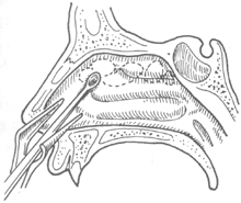

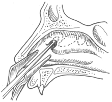
Figure 2: Curette entering the ethmoid sinus
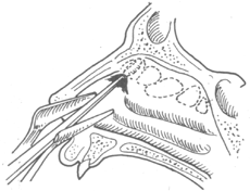
Figure 3: Scraping the anterior ethmoid sinus
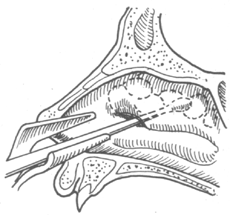
Figure 4: Scraping the posterior ethmoid sinus
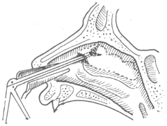
Figure 5: Removing diseased tissue within the sinus
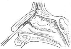
Figure 6 Enlargement of the nasofrontal duct
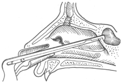
Figure 7 Enlargement of the Sphenoid Sinus Opening
III. External Ethmoidectomy
1. Indications
① Well-developed paranasal sinuses on X-ray, with sphenoethmoidal cells or maxillary ethmoidal cells; ② Chronic ethmoiditis complicated by frontal sinusitis; ③ Ethmoid sinus mycosis; ④ Foreign bodies in the ethmoid sinus; ⑤ Tumors of the ethmoid sinus; ⑥ Ethmoiditis with intraorbital or intracranial complications; ⑦ Repair of traumatic cerebrospinal fluid rhinorrhea.
2. Surgical Procedure
(1) Position and Anesthesia: The patient is placed in a supine position. Nasal mucosal surface anesthesia is applied, followed by subcutaneous infiltration anesthesia with 1–2% procaine (with a small amount of adrenaline added) along the incision. The anesthesia is extended 2 cm deep under the bony lamina of the medial orbital wall to block the anterior ethmoidal nerve.
(2) Incision: A curved incision approximately 2.5 cm long is made from the inferior edge of the eyebrow between the inner canthus and the midline of the nasal root to the infraorbital margin.
(3) Dissection: Subcutaneous tissues are dissected along the curvature of the bony wall to expose the frontal process of the maxilla and part of the nasal bone. The lacrimal bone and the lamina papyracea of the ethmoid bone are then exposed. The periosteum is incised, and the bone surface is dissected and exposed, taking care to protect the medial canthal ligament and the lacrimal sac.
(4) Opening: The ethmoid cells are entered by chiseling through the lacrimal bone. The lacrimal bone, part of the frontal process of the maxilla, and the lamina papyracea are removed to enlarge the opening of the anterior ethmoid sinus. Under direct vision, the bony septa of the air cells are removed, and all diseased tissues within the ethmoid sinus are cleared. Since the roof of the ethmoid sinus and the lamina papyracea are clearly visible, injury is unlikely. The middle turbinate should be preserved. Extending the procedure posteriorly allows visualization of the anterior wall of the sphenoid sinus and its opening, which can be explored if necessary.
(5) Packing and Suturing: After completing the debridement and hemostasis, the surgical cavity and nasal cavity are packed with antibiotic-soaked gauze strips. The incision is closed in two layers, followed by pressure dressing.
(6) Postoperative Care: The packing is removed on the second postoperative day, and sutures are removed on the sixth day.
3. Surgical Considerations
Radical intranasal ethmoidectomy requires thorough opening of all air cells while avoiding injury to adjacent organs and tissues to prevent complications, which presents certain challenges. Therefore, it is essential to pay attention to the following key anatomical landmarks during surgery.
(1) Liu Pure Brightness (5th solar term), Zhu Shijie, and others consider the middle turbinate an important landmark for intranasal ethmoidectomy. It is attached to the medial surface of the upper part of the ethmoid labyrinth, hanging between the nasal septum and the ethmoid air cells. The surgery is performed between the middle turbinate and the lamina papyracea. The average distance from the anterior end of the middle turbinate to the sphenoid sinus ostium is 34 mm, which can serve as the anterior-posterior boundary for ethmoidectomy. The average distance from the midpoint of the inferior edge of the middle turbinate to the cribriform plate is 22 mm, which can be used as a reference to avoid injury to the cribriform plate.
(2) Localization of the Lamina Papyracea: The distance between the bilateral lamina papyracea is narrower superiorly and wider inferiorly (the average superior margin is 24 mm anteriorly, 26 mm in the middle, and 28 mm posteriorly; the average inferior margin is 32 mm anteriorly, 35 mm in the middle, and 37 mm posteriorly). The lamina papyracea appears trapezoidal on the coronal plane of the ethmoid bone, located on the vertical plane of the lateral nasal wall (medial wall of the maxillary sinus) or medial to it, but never lateral. Therefore, using the lateral nasal wall as a landmark during surgery can prevent injury to the lamina papyracea or accidental entry into the orbit, damaging important nerves and blood vessels.
(3) The anterior nasal spine can serve as an external anatomical landmark. The average distance from the spine to the optic foramen is 70 mm, and the angle between the line connecting these two points and the midline averages 11.7°, which can indicate the optimal range for ethmoidectomy.
The ethmoid sinus exhibits significant anatomical variations, so the above data are for reference only.
IV. Transmaxillary Ethmoidectomy
This procedure is a multi-sinus surgery primarily involving the maxillary and ethmoid sinuses. Its advantages include the ability to treat multiple sinus infections in a single operation, no facial scarring, fewer surgical complications, and greater safety compared to intranasal ethmoidectomy. The drawback is that the anterior ethmoid air cells are less likely to be completely removed.
1. Indications
Chronic sinusitis or polyps confirmed by sinus X-ray or maxillary sinus puncture, accompanied by chronic suppurative maxillary sinusitis.
2. Surgical Procedure
First, the maxillary sinus radical operation is routinely completed. After thorough hemostasis, the inner wall is chiseled open slightly lateral to the postero-supero-medial angle of the sinus to enter the ethmoid sinus. Using a curette or round-headed biting forceps, the lesions within the ethmoid sinus are gradually cleared. The curette is then used to expand laterally to locate the lamina papyracea as a surgical landmark. Continuing posteriorly with the curette, the anterior wall of the sphenoid sinus can be reached and explored if necessary. Lesions near the openings of the anterior ethmoid air cells and the nasofrontal duct can be cleared via the nasal approach.
V. Bilateral Maxillary-Ethmoid Sinus Radical Operation via External Nasal Flip Approach
This method was first introduced by Maniglva in 1959. It allows the opening of bilateral ethmoid sinuses and the performance of a maxillary sinus radical operation in a single procedure. It offers advantages such as an expanded surgical field, clear visibility, thorough lesion removal, reduced number of surgeries, and no facial scarring.
1. Surgical Procedure
(1) Anesthesia: Same as the Caldwell-Luc operation.
(2) Incision: Along the upper labial gingival groove, cut from the midline to both sides up to the third molar, approximately 6 cm in length.
(3) Exposure of the piriform aperture and incision of its mucous membrane. The periosteum is elevated upward, laterally reaching the transition between the anterior and posterior walls of the maxilla, superiorly approaching the infraorbital foramen, and medially exposing the edge of the piriform aperture. At the midline, the medial crus of the greater alar cartilage is dissected. At the edge of the piriform aperture, the nasal cavity mucous membrane is incised from the nasal floor and cut upward along the edge of the piriform aperture to the lower border of the nasal bone.
(4) Full-thickness incision of the nasal septum: Insert the inferior turbinate scissors below the greater alar cartilage at the separation point from the nasal spine, and cut upward through the full thickness of the nasal septum up to the perpendicular plate of the ethmoid bone, ensuring a clean, single cut.
(5) Exposure of the nasal cavity roof and nasal cavity clearance: Use retractors to lift the nasolabial and cheek soft tissues along with the upper part of the nasal septum upward, fully exposing the entire piriform aperture. Part of the frontal process of the maxilla is removed with biting forceps, revealing the anterior roof of the nasal cavity and the middle and inferior turbinates. If nasal polyps or polypoid changes in the middle turbinate are present, they should be removed.
(6) Opening the ethmoid sinus: Use an ethmoid sinus curette to press the ethmoid cells at the agger nasi or ethmoid bulla, clearing all ethmoid cells from front to back and top to bottom. Suction is used to remove blood and bone fragments. The middle turbinate without polypoid changes can be preserved or pressed medially and superiorly as an anatomical landmark. The posterior ethmoid sinus opening is completed following the principles of intranasal ethmoidectomy.
(7) Clearing the maxillary sinus: Chisel open the exposed canine fossa within the surgical field, and conventionally open the anterior wall of the maxillary sinus. Depending on the extent of mucosal lesions in the sinus, partial or complete stripping and removal are performed, along with creating a medial wall antrostomy. The posterior ethmoid air cells can be opened and cleared posterior to the superomedial angle of the maxillary sinus, coordinated with intranasal ethmoidectomy to ensure no residual infection remains.
(8) After completing the surgery, reposition the nasolabial soft tissues. The mucous membrane at the piriform aperture does not require suturing. The nasal septum incision is repositioned and aligned, fixed bilaterally with nasal packing without suturing. Only a few sutures are needed at the labial gingival groove incision. Pressure dressing and postoperative care are the same as the Caldwell-Luc operation. Sutures are removed on the 6th day.
VI. Functional Endoscopic Ethmoidectomy
This surgical technique was innovated in 1978 by Messerklinger, who synthesized previous experiences and theorized it as a new rhinological procedure, later refined and popularized by Kennedy, Stammberger, and others. The goal of this surgery is to transform traditional radical (destructive) procedures into functional (reconstructive) operations, thereby completely curing sinusitis and restoring its original functions. According to the characteristics of nasal airflow, inhaled air first impacts the middle turbinate, middle meatus, and anterior ethmoid sinuses upon entering the nasal cavity, making this area most susceptible to infection and allergen attacks. Modern theory suggests that the ethmoid sinuses have the highest incidence of disease and serve as the origin of pathologies in other sinuses. Therefore, the focus of functional endoscopic sinus surgery (FESS) lies in ethmoid sinus surgery. Previously, conditions such as irreversible maxillary, frontal, or sphenoid sinusitis were thought to require separate interventions. However, by surgically clearing lesions in the anterior ethmoid sinuses and restoring normal sinus ventilation and drainage, inflammation in other sinuses can gradually resolve without additional surgery.
1. Preoperative Preparation
(1)Instrument Preparation: Select nasal endoscopes with 0°, 30°, 70°, 90°, and 120° viewing angles, which provide strong illumination and a field of view without blind spots. Prepare several ethmoid forceps with various curvatures, one straight and one angled suction device, a set of nasal septum surgical instruments, one nasal snare, one nasal knife, one syringe and a long needle (No. 5), one nasal forceps, one pair of scissors, one nasal speculum, one electrocoagulation hemostat, and one television and video recording system.
(2)Patient Preparation
① Inquire about any history of nasal surgery, with particular attention to those with a history of nasal polyp surgery. Patients taking salicylic acid preparations should postpone the surgery.
② Conduct a comprehensive systemic examination, including routine hematuria tests and an electrocardiogram.
③ Perform an eye examination, paying attention to visual acuity, visual field, intraocular pressure, extraocular muscle function, and degree of exophthalmos.
④ Nasal preparation: Trim nasal hair, perform negative pressure displacement, and administer antibiotic nasal drops.
⑤ Prepare 500ml of blood; for repeat surgeries, additional blood should be prepared.
⑥ Take X-rays and CT scans of the nasal sinuses, paying attention to the condition of the ethmoid sinus roof and the lamina papyracea.
⑦ Begin bacterial culture and drug sensitivity testing of nasal secretions several days before surgery. Since anaerobic bacteria are often pathogenic in sinusitis, anaerobic culture is essential. If positive, administer metronidazole 200mg three times daily for two days before surgery.
⑧ Provide thorough explanations to the patient and their family, objectively analyzing and estimating surgical outcomes and potential complications. Especially when the surgery involves the anterior skull base, sphenoid sinus, or orbit, emphasize the inherent risks. Do not overlook obtaining signed surgical consent.
⑨ Administer 0.1g of Luminal intramuscularly 30 minutes before surgery.
2. Surgical Position and Anesthesia
(1)Position: Place the patient in a supine or 30° reclining position.
(2)Anesthesia: For local anesthesia in bilateral sinus surgery, use 25ml of 2% dicaine mixed with 2–3ml of 0.1% adrenaline. Soak cotton pads in the solution, wring them to Grade I dryness, and apply them to the nasal mucosa twice, with a 5-minute interval between applications. For the middle turbinate and agger nasi, use a No. 5 long needle to perform submucosal infiltration anesthesia with 5ml of 1% lidocaine mixed with 2–3 drops of 0.1% adrenaline. For general anesthesia with tracheal intubation, the nasal mucosa should still be treated with adrenaline-soaked cotton pads to reduce intraoperative bleeding.
3. Surgeon’s Position and Responsibilities
The surgeon stands on the patient’s right side. The first assistant stands beside the surgeon and is responsible for instruments, dressings, anesthesia, and other tasks directly related to the surgery. The second assistant stands on the patient’s left side and manages the video monitoring system, capturing photos and videos as directed by the surgeon. One circulating nurse is responsible for IV fluids, blood transfusions, injections, and providing any surgical supplies requested by the surgeon.
4. Surgical Procedure
(1)Facial Disinfection: Use 75% alcohol for facial disinfection and merbromin for the nostrils. Avoid thimerosal, as it can damage the mucosa. When draping, ensure the patient’s eyes remain uncovered for continuous monitoring of vision and extraocular muscle function during surgery.
(2)Incision: If there are no polyps in the middle meatus, make a longitudinal incision anterior to the middle meatus, corresponding to the anterior edge of the middle turbinate, or a crescent-shaped incision at its anteroinferior edge. If polyps are present in the middle meatus or the middle turbinate has polypoid changes, make an incision between the medial surface of the middle turbinate and the polyp. A laser knife can minimize bleeding, and a 0° endoscope can guide the procedure.
(3)Use a nasal septum dissector to separate the middle meatal mucosa and expose the ethmoid bulla. Gently press or use straight forceps to open the ethmoid bulla. For thick bone walls, a chisel may be used. To widen the surgical pathway, the middle turbinate can be pushed toward the nasal septum. The size of the ethmoid bulla can be referenced from preoperative imaging studies.
(4) During the cleaning of the ethmoid sinus, the roof of the ethmoid sinus appears pale yellow under the endoscope. Special care should be taken when operating in this area, typically using a 30-degree or 70-degree scope with a curette, avoiding the use of polyp forceps.
(5) Clearing the anterior ethmoid cells and supraorbital ethmoid cells Using a 70-degree endoscope with a large-opening polyp forceps, clear the anterior ethmoid cells and supraorbital ethmoid cells, extending superiorly to the floor of the frontal sinus, laterally to the lamina papyracea (which is continuous with the lamina papyracea of the middle ethmoid region), and anteriorly to the frontal process of the maxilla. Occasionally, the anterior ethmoid artery pulse may be observed along the skull base; care must be taken to avoid injury. When clearing the anterior ethmoid cells, be cautious not to injure the lacrimal sac or nasolacrimal duct.
(6) Clearing the posterior ethmoid cells Using a 4mm 0-degree wide-angle endoscope with a large-opening straight forceps, switch to an opening straight forceps when entering the most posterior ethmoid cells. Remove all posterior ethmoid cells, extending superiorly to the ethmoid roof, laterally to the lamina papyracea, posteriorly to the anterior wall of the sphenoid sinus, and medially to the middle turbinate, creating a single cavity within the entire ethmoid sinus.
(7) Opening and exploring the frontal sinus Using a 70-degree endoscope with a curette or suction, explore the floor of the frontal sinus. After locating the frontal sinus ostium, use the curette to enlarge the opening circumferentially. A bony prominence exists between the frontal recess and the anterior ethmoid roof, serving as an important landmark. The area anterior to this prominence contains the frontal sinus floor and ostium, while the area posterior to it is the ethmoid roof (i.e., the anterior skull base), where no manipulation should be performed. The enlarged frontal sinus ostium should measure at least 0.5cm to ensure adequate drainage and prevent postoperative stenosis. Unless polyps or neoplasms are present, the mucosal lining of the frontal sinus generally does not require intervention.
(8) Opening and exploring the maxillary sinus Under the guidance of a 70- or 90-degree endoscope, use a reverse-cutting forceps to enlarge the maxillary sinus ostium to approximately 1.0cm. Inspect the sinus with endoscopes of varying angles. If polyps or cysts are found, remove them. Thickened mucosa may be left untreated. If purulent secretions are abundant, create an additional opening in the inferior meatus to establish two ostia for improved ventilation and drainage. This method is also referred to as combined antrostomy.
(9) Opening and exploring the sphenoid sinus After clearing the posterior ethmoid cells, if the sphenoid sinus ostium is low-lying, enlarge it circumferentially with a curette. If the ostium is high-lying, use sharp ethmoid forceps to fracture the anterior wall of the sphenoid sinus, confirm its position with a probe, and then enlarge it with forceps. The distance from the anterior wall of the sphenoid sinus to the anterior naris is typically 7.5–7.8cm, rarely less than 7.2cm, which can serve as a reference for locating the anterior wall. According to Xu Geng’s observation of 100 adult skulls, approximately 20% have sphenoethmoid cells (Onodi cells), which should not be mistaken for the sphenoid sinus to avoid complications. Preoperative coronal CT scans of the sinuses can guide the surgery. If uncertainties arise during the procedure, a 0-degree endoscope or macroscopic inspection after withdrawing the endoscope may be used.
After completing the entire procedure, irrigate the ethmoid sinus with saline and inspect the cavity for residual diseased mucosa or bone fragments, removing them as needed. Scrape away intercellular bony septa and smooth the edges of all opened sinus ostia. For active bleeding, use bipolar electrocautery for hemostasis. Finally, lightly pack the surgical cavity with gelatin sponge or Vaseline gauze.
5 Postoperative Management
(1) For patients under local anesthesia, place them in a semi-sitting position postoperatively. Monitor for posterior nasal bleeding and instruct the patient to spit blood into a kidney basin. Minor oozing may not require intervention, but significant bleeding warrants repacking.
For patients under general anesthesia, ensure airway patency before awakening, frequently suctioning oropharyngeal secretions and blood. On the second postoperative day, they may ambulate, with management otherwise similar to that for local anesthesia patients.
(2) Administer intravenous 5% or 10% glucose solution, with 4–6g of cephalosporin per 500ml. Provide a semi-liquid diet.
(3) Routinely examine the eyes, including eyelids, bulbar conjunctiva, extraocular muscles, intraocular pressure, visual acuity, visual fields, and proptosis, comparing findings with preoperative status. Grade I eyelid congestion and edema are common postoperatively due to impaired periorbital venous drainage and typically resolve after gauze removal. If bulbar conjunctival congestion, restricted eye movement, decreased vision, or proptosis occurs, orbital involvement is suggested, requiring immediate gauze removal and prompt intervention.
(4) The routine time for removing the gauze strip is 1–2 days after surgery. Pay attention to whether there is cerebrospinal fluid rhinorrhea. If cerebrospinal fluid rhinorrhea occurs, the patient is prohibited from blowing their nose, and sufficient antibiotics should be used to prevent intracranial infection. Rinse the surgical cavity daily with physiological saline containing antibiotics for 5–7 consecutive days.
(5) Cleaning the Surgical Cavity This task is a long-term process crucial to the success of the surgery and must be performed under endoscopy. It is divided into three stages.
① Early Postoperative Period (7–10 days): Daily suction of blood clots from the cavity is required. Residual diseased tissue should be removed with polyp forceps, and the nasal mucosa should be contracted using 1% Ephedrine-soaked cotton pads. Special attention must be paid to the openings of the anterior ethmoid sinus and the front part of the middle meatus. If the cavity becomes obstructed and fails to drain, the surgery is deemed a failure. New granulation tissue or scabs may form postoperatively and should also be thoroughly removed, followed by saline irrigation. The scabs typically resolve within 2–3 weeks.
② Within 3 Months Postoperatively: Follow-up visits should be scheduled every 1–2 weeks. The cavity should be contracted, cleaned, and irrigated as described above. Monitor for secondary infections, polyp recurrence, sinus ostium narrowing, or middle meatus adhesions, and address them promptly to facilitate the restoration of ciliary function in the surgical cavity.
③ Within 6 Months Postoperatively: Follow-up visits should be scheduled every 1–2 months, with the same treatment approach as before to consolidate surgical outcomes. Generally, anterior rhinoscopy should reveal a normalized middle turbinate and a smooth, unobstructed middle meatus. If endoscopy detects sinus ostium obstruction or adhesions, a second surgery may be necessary. However, due to the altered nasal anatomical landmarks, the second surgery is more challenging and is best performed by the original surgeon.
Common Complications of Ethmoid Sinus Surgery In sinus surgery, ethmoid sinus surgery is the most prone to complications because the ethmoid sinus is the smallest among all the paranasal sinuses. According to Rice (1989), a normally developed ethmoid sinus measures 4–5 cm in length, with the anterior ethmoid being 2.5 cm in height, 0.5 cm in width, the posterior ethmoid 1.5 cm in width, and the ethmoid roof 0.3 cm in width. Performing surgery within such a confined space increases the risk of injuring the lateral lamina papyracea or the cribriform plate at the roof of the ethmoid sinus if bleeding occurs or lighting is inadequate. Among the various ethmoidectomy techniques, intranasal ethmoidectomy is the most likely to cause intraorbital and intracranial complications, followed by the Lima procedure, while external ethmoidectomy carries the lowest risk. In recent years, functional endoscopic sinus surgery (FESS) has been introduced, but despite advanced equipment, complications have been reported both domestically and internationally due to surgeons' limited experience.
1. **Intracranial Complications** — Life-threatening.
(1) **Injury to the skull base** — Cerebrospinal fluid rhinorrhea, intracranial pneumatosis (pneumocephalus), often caused by surgical instruments being directed too upward, surpassing the attachment of the middle turbinate or appearing higher than the inner canthus plane.
(2) **Intracranial hemorrhage** — Subdural hematoma, frontal lobe hematoma, or carotid-cavernous fistula, resulting from excessive upward forceps extraction of tissue at the ethmoid roof or injury to blood vessels while removing tissue from the lateral wall of the sphenoid sinus. If the internal carotid artery ruptures, the patient may rapidly die from epistaxis.
(3) **Intracranial infection** — Common conditions include meningitis, subdural abscess, and brain abscess.
2. **Orbital and Ocular Complications** — Risk of blindness.
Main manifestations include proptosis, visual impairment, ophthalmoplegia (strabismus, diplopia), and epiphora. Causes include:
(1) **Lamina papyracea injury** — Mild cases may present with orbital emphysema or subcutaneous ecchymosis of the eyelid, while severe cases involve rupture of the anterior ethmoidal artery, leading to orbital hematoma and rapid blindness. Injury to the medial rectus muscle may cause outward strabismus and diplopia.
(2) **Optic nerve injury** — Includes injury to the optic canal or retrobulbar segments. If the extracted tissue contains yellow, soft orbital fat, the optic nerve, orbital vessels, or medial rectus muscle may be damaged, potentially causing reflexive retinal artery spasm or embolism, leading to blindness.
(3) **Lacrimal system injury** — Involving the lacrimal sac or nasolacrimal duct, with epiphora as the primary sign.
(4) **Orbital infection** — Conditions such as orbital periostitis, orbital cellulitis, or orbital abscess may develop days post-surgery, with fever and eye pain as primary symptoms.
3. **Intranasal Complications**
(1) **Hyposmia or anosmia** — Often due to excessive loss of olfactory mucosa.
(2) **Nasal and sinus adhesions** — Adhesions at the osteomeatal complex may lead to failure of functional endoscopic sinus surgery.


