
| disease | Thoracic Outlet Syndrome |
Thoracic outlet syndrome is a series of symptoms caused by compression of the subclavian artery, vein, and brachial plexus at the superior thoracic aperture.
bubble_chart Etiology
The causes of nerve and/or vascular compression include abnormal bone structures, such as cervical ribs, an elongated transverse process of the 7th cervical vertebra, malformations of the first rib or clavicle, exostoses, fractures of the clavicle or first rib due to trauma, dislocation of the humeral head, and other conditions. Additionally, spasms or fibrosis of the scalene muscles, drooping shoulders due to leukorrheal disease, and excessive abduction of the upper limbs can all lead to narrowing of the thoracic outlet, resulting in compression symptoms of the subclavian vessels and brachial plexus. Furthermore, normal movements of the upper limbs, such as abduction of the arm, drooping of the shoulders (to be decocted later), neck extension, turning the face to the opposite side, and deep inhalation, can also reduce the costoclavicular space, exacerbating the compression of nerves and blood vessels (Figure 1).
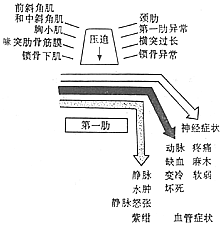
Figure 1 Schematic diagram of the pathogenesis of thoracic outlet syndrome—scissor-like compression.
The superior thoracic aperture is bounded superiorly by the clavicle, inferiorly by the first rib, anteriorly by the costoclavicular ligament, and posteriorly by the middle scalene muscle. The aforementioned costoclavicular space is further divided into anterior and posterior parts by the anterior scalene muscle. The subclavian vein lies between the anterior scalene muscle and the subclavius muscle anteriorly, while the subclavian artery and brachial plexus are located between the anterior and middle scalene muscles posteriorly (Figure 1).
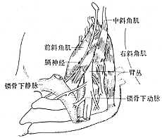
Figure 1 Anatomy of the scalene muscles in relation to the subclavian vessels and brachial plexus (with clavicle removed)
bubble_chart Pathological Changes
Nerve compression injury often presents as pseudo-inflammatory swelling, with sensory fibers being the first to be affected, while motor nerves only show compression in advanced stages. This symptom is severe and difficult to recover from. Prolonged nerve compression can lead to vasomotor dysfunction through the sympathetic nerves. The vascular wall of the subclavian stirred pulse may undergo changes, including thickening of the stirred pulse outer membrane, interstitial edema, and thickening of the same membrane accompanied by intraluminal thrombosis. Early thrombosis is of the fibrin-platelet type and may manifest as Raynaud's phenomenon. The vasoconstrictive reflex of sympathetic nerve fibers can exacerbate vascular obstruction in the fingertips. When veins are compressed during excessive abduction or adduction, retrograde blood stasis and elevated peripheral venous pressure can be observed, which return to normal once the compression is relieved. Repeated injury to the venous wall can lead to post-inflammatory fibrotic-like changes, causing the veins to appear white, lose their semi-translucent state, and significantly narrow in diameter, forming collateral circulation. The early trend is venous thrombosis, and if collateral circulation has not yet developed, it may result in necrosis of the fingertips.
bubble_chart Clinical ManifestationsIt is divided into two categories: nerve compression and vascular compression. Symptoms of nerve compression are more common, and there are also cases where both nerves and blood vessels are compressed simultaneously.
(1) Symptoms of nerve compression include pain, paresthesia, and numbness, often occurring in the ulnar nerve distribution area of the fingers and hand. Pain may also radiate to the upper limb, shoulder girdle, and ipsilateral shoulder and back. In advanced stages, there may be loss of sensation, motor weakness, atrophy of the thenar and interosseous muscles, and paralysis of the extensor muscles of the 4th and 5th fingers, leading to claw hand deformity.
(2) Symptoms of stirred pulse compression include ischemic pain, numbness, fatigue, paresthesia, coldness, and weakness in the arm or hand. Compression of the stirred pulse can lead to distal dilation and thrombosis, causing distal ischemia. Venous compression may result in pain, swelling, aching pain, distal swelling, and cyanosis.
bubble_chart Auxiliary Examination
First, it is determined that nerve compression mostly occurs in the ulnar nerve distribution area. Compression of the stirred pulse may involve weakened or absent radial stirred pulse and brachial stirred pulse, with murmurs heard in the supraclavicular and axillary regions. Venous compression manifests as venous distension, distal edema, and cyanosis. The following examination methods are helpful for diagnosis.
1. Upper Limb Abduction Test The upper limb is abducted to 90°, 135°, and 180° with the hand externally rotated and the neck extended. This compresses the subclavian neurovascular bundle beneath the insertion of the pectoralis minor muscle and between the clavicle and the first rib, causing or exacerbating pain in the neck, shoulder, and upper limb. The radial stirred pulse weakens or disappears, blood pressure drops by 2.0 kPa (15 mmHg), and a systolic murmur is heard in the subclavian stirred pulse region (Figure 1).
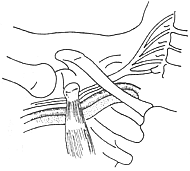
Figure 1: The affected upper limb is abducted beyond 90°, the hand is externally rotated, and the neck is extended, compressing the subclavian artery, vein, and brachial plexus beneath the insertion of the pectoralis minor muscle and between the clavicle and the first rib.


Figure 2: Adson Test
3. Ulnar Nerve Conduction Velocity Measurement The ulnar nerve conduction velocity is measured separately at the thoracic outlet, elbow, and forearm. Normal values are 72 m/s at the thoracic outlet, 55 m/s at the elbow, and 59 m/s in the forearm. In thoracic outlet syndrome patients, the ulnar nerve conduction velocity at the thoracic outlet decreases to 32–65 m/s, averaging 53 m/s.
4. Doppler Ultrasound and Photoelectric Flowmetry These serve as methods to assess vascular compression in thoracic outlet syndrome but are not specific. However, they can exclude vascular diseases. Preoperative and postoperative blood flow assessments help evaluate surgical efficacy.
5. Selective Angiography Used for severe arterial or venous compression, combined with stirred pulse aneurysms, atherosclerotic patches, embolisms, or venous thrombosis to clarify the nature of the lesion and exclude other vascular pathologies.
Based on medical history, local physical examination, chest and cervical spine X-rays, and ulnar nerve conduction velocity testing, a definitive diagnosis can generally be established. The differential diagnosis of thoracic outlet syndrome should consider cervical spine disorders, brachial plexus or peripheral nerve diseases of the upper limb, vascular diseases, and heart, lung, or mediastinal diseases. For cases suspected of having colicky heart pain, electrocardiography and selective coronary angiography should be performed.
bubble_chart Treatment Measures
It can be divided into conservative treatment and surgical treatment.
(1) Conservative treatment: Suitable for patients with mild symptoms or initial onset. Methods include:
1. Inject 5ml of 1% procaine plus 1ml of hydrocortisone into the tender area of the left or right supraclavicular fossa, administered intramuscularly once a week for 3–5 sessions as one course. This is notably effective for those with a history of local muscle strain.
3. Physical therapy: Apply diathermy or iodine iontophoresis to the supraclavicular fossa.
4. Physical exercises for shoulder girdle muscles and cervical traction.
(2) Surgical treatment: Suitable for patients whose symptoms do not improve or even worsen after 1–3 months of non-surgical treatment, with ulnar nerve conduction velocity below 60m/s through the thoracic outlet; angiography showing significant stenosis or obstruction of the subclavian stirred pulse and veins; or those with severe local pain or pronounced venous compression symptoms.
The surgical principle is to relieve the bony scissor-like compression on the neurovascular bundle. The entire first rib must be resected, and related compressive factors must be eliminated to allow the brachial plexus and subclavian stirred pulse to descend without causing deformities or complications.
There are two surgical approaches:
(1) Axillary approach: Under general anesthesia or high epidural anesthesia, the patient is placed in an oblique position with the affected limb elevated at 45°. After raising the upper limb, a 6–7cm transverse incision is made at the level of the third rib below the axillary hairline. Dissection is performed between the pectoralis major and latissimus dorsi muscles to reach the thorax, followed by upward separation beneath the fascial membrane to the apex of the axilla. The neurovascular bundle is visualized at the upper edge of the first rib. Raising the upper limb displaces the neurovascular bundle away from the first rib. The anterior scalene muscle is severed, and the first rib and periosteum are resected, extending anteriorly to the costal cartilage and posteriorly to the transverse process. Postoperatively, the bone stump is examined for any compression on the brachial plexus. This approach is less traumatic and involves less bleeding, but the exposure is poor, and incomplete resection of the first rib may occur (Figure 1).
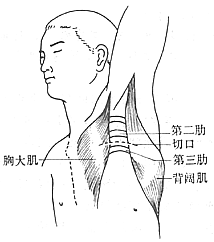
Figure 1: Surgical incision for thoracic outlet syndrome via the axillary approach.
The patient is placed in an oblique lateral position with the upper limb raised, and the incision extends from above the third rib to below the axillary hairline.
(2) Parascapular approach: Under general anesthesia, the patient is placed in a lateral position with the affected limb raised 90°. The incision starts from the high parascapular region, follows the medial border of the scapula downward, and curves toward the axilla. The latissimus dorsi, water calptrop base peel-shaped muscle, and serratus anterior are severed. The scapula is retracted upward and outward, and the fibers of the middle scalene muscle are cut to expose the first rib. The posterior segment of the second rib is resected to enhance exposure of the first rib and decompress the second intercostal nerve. This also enlarges the thoracic apex space in cases of cervical scoliosis or conical chest. The anterior scalene muscle and the entire first rib are severed, and any bony abnormalities such as cervical ribs, elongated transverse processes of vertebral bodies, or abnormal fibrous bands should be resected. This approach involves a larger incision, and meticulous hemostasis is required postoperatively to prevent hematoma-induced adhesions. It allows satisfactory resection of the first rib and elimination of related compressive factors, making it suitable for reoperation cases. The drawbacks include greater trauma and more bleeding. Surgical complications may include injury to the pleural membrane causing pneumothorax, intraoperative traction on the brachial plexus leading to arm numbness or weakness, or postoperative hematoma infection. Symptoms resolve in over 90% of cases postoperatively (Figure 2).
|
|
(1) Schematic of the posterolateral incision. | (2) Free anterior bone membrane |
|
|
(3) Resection of the first rib near the transverse process | (4) Free the anterior periosteum |
| |
(5) After rib resection, remove the periosteum and attached scalene muscles | |
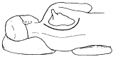
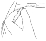
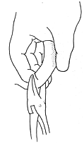
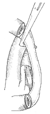

Figure 2
bubble_chart Other Related Items


