| disease | Chronic Cervicitis |
| alias | Chronic Cervicitis |
Chronic cervicitis is one of the most common gynecological diseases, which may occur after acute cervicitis or due to cervical laceration caused by various reasons leading to deformation of the cervical os, making it highly susceptible to external bacterial infections.
bubble_chart Etiology
Garden Balsam Seed Cervicitis tends to become chronic mainly because the cervical mucosa has numerous folds and the glands are grape-like, making it extremely difficult to eradicate pathogens once they invade the deeper parts of the glands. This leads to recurrent episodes, prolonged duration, and the development of a chronic sexually transmitted infection. Similar to how conditions like tonsillitis, sinusitis, or dental caries can cause inflammation in other parts of the body, such as arthritis or muscle rheumatism, and affect overall health.
bubble_chart Pathological Changes
Chronic cervicitis can manifest in various local forms:
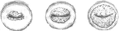
Cervical erosion (cervical erosion) Cervical erosion is the most common local feature in the inflammatory and sexually transmitted disease progression of chronic cervicitis. The cervical surface appears as a red lesion, resulting from the shedding of squamous epithelium and its replacement by columnar epithelium, exposing the underlying blood vessels. The eroded area has a clear boundary with the surrounding normal squamous epithelium. Since it is not a true erosion, it is also called "pseudoerosion." Depending on the severity of inflammation and the growth rate of columnar epithelium, cervical erosion can present in three types.
(1) Simple type: In the initial stage of inflammation, the eroded area is covered by a single layer of columnar epithelium, presenting a flat and smooth surface.
(2) Granular type: Due to excessive proliferation of cervical glandular epithelium and stromal hyperplasia, the eroded surface becomes uneven and granular.
(3) Papillary type: Significant hyperplasia of glandular epithelium and stroma leads to a more pronounced uneven surface, forming papillary protrusions.
Clinically, the extent of erosion is often classified as mild, moderate, or grade III. Erosion covering less than one-third of the total cervical area is classified as grade I (Ⅰ°), one-third to one-half as grade II (Ⅱ°), and more than half as grade III (Ⅲ°) (Figure 1).
Grade I Grade II Grade III
Figure 1 Cervical erosion
Particularly noteworthy is the healing process of cervical erosion. As the inflammatory condition slightly subsides, the adjacent squamous epithelium grows beneath the columnar epithelium covering the eroded area, gradually displacing the glandular epithelium until the area is completely covered by squamous epithelium and healed. Although direct replacement by squamous epithelium occurs, indirect replacement is more common. That is, beneath the columnar epithelium, there is often a row of smaller round cells called basal cells or reserve cells. During the healing process of erosion, these cells proliferate and eventually differentiate into squamous epithelium. Healing often occurs in patches, and because this newly formed squamous epithelium grows on an inflammatory tissue base, the superficial cells are prone to shedding and thinning, easily reverting to erosion with minor irritation. Thus, healing and inflammatory extension alternate, making complete recovery difficult without thorough treatment.
The aforementioned healing process not only occurs on the surface of the erosion but also affects the columnar epithelium covering the recessed glands and hyperplastic glandular spaces, which can similarly be replaced by stratified epithelium. This stratification and epidermization of glandular epithelium is commonly referred to as "squamous metaplasia." The extent of metaplasia varies greatly; sometimes the entire glandular epithelium is replaced, sometimes only one side or the opening of the gland, and in some cases, the entire glandular structure forms solid cell clusters within the cervical stroma. Due to the high incidence of chronic cervicitis, squamous metaplasia is found in 70-80% of cervical biopsies. Squamous metaplasia is a change during the healing process of erosion and does not indicate a tendency to form cancer. It is not a precancerous lesion and should not be confused with anaplasia or atypical hyperplasia.
In addition, the formation of cervical erosion may also be attributed to the following two reasons: ①So-called congenital erosion: During the late stage of embryonic development [third stage], both the vagina and the vaginal portion of the uterine cervix are covered by transitional epithelium. By the sixth month, this epithelium extends into the cervical canal. At full term, the columnar epithelium of the cervical mucous membrane grows outward, surpassing the external os of the uterus. Approximately one-third of newborn female infants retain this condition, which resembles the inflammatory cervical erosion seen in adults, hence the term "congenital cervical erosion." However, this condition typically persists for only a few days and naturally regresses once the maternal estrogen levels decline. ②Cervical erosion caused by the proliferation of columnar epithelium from the cervical mucous membrane, extending beyond the external os of the cervix. This appears similar to inflammatory cervical erosion but occurs only during the reproductive age when ovarian function is robust, not during adolescence or after menopause. It is also more common during pregnancy and often tends to resolve spontaneously postpartum. Although patients may experience increased leucorrhea, it is clear and mucous. Pathological examination reveals no inflammatory cell infiltration beneath the columnar epithelium or only a few lymphocytes, characterized by the aforementioned papillary and glandular erosion tissue patterns. All these phenomena suggest that the formation of such erosion may be due to an imbalance in sex hormones rather than inflammation, although inflammation may secondarily develop on the basis of erosion. However, this is merely a consequence and not the cause of erosion. Erosion may result from the effects of estrogen, but some animal experiments have shown that injecting testosterone produces changes similar to human glandular erosion. Therefore, it is believed that androgens can transform uterine cervical epithelium into a mucous type and promote gland formation. Progesterone has effects similar to androgens in this regard, while estrogen causes epithelial hyperplasia, leading to highly keratinized stratified squamous epithelium.
In summary, the disease causes of cervical erosion are mostly inflammation, but may also be caused by endocrine disorders. In differential diagnosis, attention should be paid to the onset period and the presence of predisposing factors and signs related to inflammation. Pathological examination can also serve as a reference, and the treatment varies slightly (see the treatment section for details).In chronic cervicitis, the cervical glands and surrounding tissues undergo hyperplasia. When the glandular ducts are compressed by the surrounding tissues, the glandular openings become blocked, preventing the secretions within the glands from flowing out and leading to retention. This causes the glandular cavities to expand, forming cystic masses of varying sizes, known as "retention cysts of the cervical glands" or Naboth's cysts (Figure 2). The contained mucus is usually clear and transparent but may become turbid or purulent due to concurrent infection. The cysts are generally small and scattered and may protrude from the cervical surface. The smaller ones are only the size of a foxtail millet grain, while the larger ones can reach the size of a corn kernel, appearing bluish-white and sometimes accompanied by erosion. However, they are also commonly found on a smooth cervical surface. Due to long-term stimulation from chronic inflammation, the cervical tissue repeatedly experiences congestion, edema, inflammatory cell infiltration, and connective tissue hyperplasia, leading to cervical hypertrophy. In severe cases, the cervix may enlarge to more than twice its normal size. After the inflammation resolves, the congestion and edema lessen or disappear. However, due to fibrosis, even though the cervix is fully covered by squamous epithelium and appears smooth, it retains its hypertrophied volume. The cervical gland cysts also maintain their protruding cystic appearance.
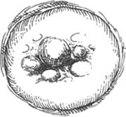
Figure 2 Cervical gland cysts
bubble_chart Clinical Manifestations
(1) Increased leucorrhea: Sometimes it is the only symptom of chronic uterus cervicitis. It is usually thick mucus or purulent mucus. Sometimes the discharge may contain blood streaks or a small amount of blood, and there may also be contact bleeding. Due to the stimulation of leucorrhea, it can cause external cutaneous pruritus.
(2) Pain: Pain often occurs in the lower abdomen or lumbosacral region, and sometimes it can appear in the upper abdomen, thighs, and hip joints. The pain worsens during menstruation, defecation, or sexual intercourse, especially when the inflammation spreads backward along the uterus sacral ligament or extends along the base of the broad ligament, forming chronic uterus connective tissue inflammation. When the main ligament of the uterus cervix thickens, the pain becomes more severe. Touching the uterus cervix immediately causes pain in the iliac fossa and lumbosacral region, and some patients may even experience nausea, affecting sexual life.(3) Bladder and intestinal symptoms: Chronic uterus cervicitis can spread through the lymphatic system or directly extend to the bladder trigone or the connective tissue around the bladder. As a result, the urge to urinate occurs as soon as there is urine in the bladder, leading to symptoms such as frequent urination or difficulty urinating. However, the urine is clear, and routine urine tests are normal. In some cases, the inflammation continues to spread or follows the lymphatic pathways connecting the uterus cervix, bladder trigone, and ureters, resulting in secondary urinary tract infections. The incidence of chronic pyelonephritis in adult women is several times higher than in men, which may be related to this condition.
Intestinal symptoms are less common than bladder symptoms, and some patients experience pain during bowel movements.
(4) Other symptoms: Such as menstrual irregularities, dysmenorrhea, pelvic heaviness, infertility, etc.
Because the symptoms of chronic cervicitis are often masked by other gynecological conditions, it is usually discovered during routine gynecological examinations. Diagnosis can be made by observing a bright red, finely granular erosion area on the cervix and purulent mucoid leucorrhea from the cervical canal through speculum examination. In some cases, the cervix may show localized congestion and hypertrophy.
bubble_chart Treatment Measures
After excluding sexually transmitted diseases and controlling specific or non-specific infections, local treatment is the main approach, allowing the columnar epithelium of the eroded area to necrotize and shed, followed by coverage with newly formed squamous epithelium.
(1) **Drug Therapy**: There are numerous drug treatment methods for chronic cervicitis, with the following being commonly used:
1. **Local Vaginal Irrigation and Medication**: This is the most commonly used treatment method. Irrigation can be performed using a 1:5000 potassium permanganate solution, 1:1000 benzalkonium bromide solution, 1% acetic acid solution, or 0.5–1% lactic acid solution. For superficial Grade I cases, a cotton swab dipped in 5–10% iodine tincture or 5–10% silver nitrate solution can be used to locally cauterize the eroded area once a week to promote healing. However, care must be taken to avoid the medication spreading to normal mucous membranes outside the affected area. After applying silver nitrate, gently wipe the area with a saline-soaked cotton ball. This method is now less commonly used. Local application of chloramphenicol and prednisone tablets (250mg chloramphenicol and 5mg prednisone per tablet) can be inserted deep into the vagina nightly or every other night, with 10 applications constituting one treatment course. The efficacy is comparable to that of general disinfectant irrigation, and the method can be selected based on individual circumstances.
2. **Vaginal Lateral Fornix Block**: For details, refer to the chapter on other therapies regarding block therapy. This method is suitable for chronic cervicitis combined with parametritis, significant lumbosacral pain, or cervical motion tenderness that severely affects sexual life.
3. **Chinese Medicinal Lotion (Jie Er Yin)**: Suitable for various acute and chronic cervicitis cases. Its main ingredients include Cnidium Fruit, Phellodendron Bark, Sophora, and Atractylodes Rhizome. Generally, a 10% solution is used for vaginal irrigation or sitz baths once daily, with two weeks constituting one treatment course.
(2) **Physical Therapy**: Currently, this is the most effective and shortest-duration treatment for cervical erosion, suitable for cases with extensive erosion and deep inflammatory infiltration. Typically, only one treatment session is required for cure.
1. **Electrocoagulation**: Previously, radial electrocoagulation (electrocauterization) was used, but the healing time was prolonged (6–8 weeks). Currently, electrocoagulation is more commonly employed to flatten the entire eroded area, hence also referred to as "electric ironing." According to a summary of 2095 cases from Shandong Medical University Affiliated Hospital, the one-time effectiveness rate of electrocoagulation for cervical erosion was 100%. **Procedure**: - Set up the electrocoagulation device and routinely disinfect the vulva, vagina, and cervix. - Use a vaginal speculum to expose the cervix and apply the electrocoagulation probe evenly over the eroded area, slightly exceeding its boundaries. - The coagulation depth should be about 0.2cm; excessive depth may cause bleeding and slower healing, while insufficient depth may reduce efficacy. - After electrocoagulation, sprinkle nitrofurazone powder or apply chlortetracycline glycerin to the wound.
2. **Cryotherapy**: This is an ultralow-temperature therapy (-196°C) using liquid nitrogen as the cooling source. An appropriate probe is selected based on the extent of erosion. To enhance efficacy, a "freeze-thaw-freeze" method can be employed: freeze for 1 minute, thaw for 3 minutes, and refreeze for 1 minute. **Advantages**: Simple operation, with rare postoperative bleeding or cervical stenosis. **Disadvantage**: Significant postoperative anal discharge.
3. Laser Therapy: This is a high-temperature treatment that can reach over 700°C. It primarily causes the necrotic tissue to carbonize and form a scab, which then falls off, leaving the wound covered by newly formed squamous epithelium. For treating cervical erosion, a carbon dioxide laser with an infrared wavelength of 10.6μm is typically used. Preparations before the treatment are the same as for electrocoagulation. Its advantages, in addition to thermal effects, include pressure, photochemical, and electromagnetic field effects. Therefore, it has therapeutic benefits such as anti-inflammatory effects (stimulating the body to produce a stronger defensive immune response), pain relief (reducing tissue edema and minimizing chemical and mechanical irritation to nerve endings), and promoting tissue repair (enhancing the anabolic metabolism of epithelial cells, stimulating epithelial proliferation, and accelerating wound healing). As a result, the treatment time is short, and the cure rate is high.
The treatment time for physical therapy should be performed within 3 to 7 days after the menstruation is clean. It is contraindicated in cases of acute genital inflammation. After physical therapy, there may be an increase in anus and vaginal secretions, so it is important to keep the vulva clean. Before the wound has healed (4 to 8 weeks after the procedure), avoid taking baths, sexual intercourse, and vagina douching. If cryotherapy is to be performed, the patient should be asked about any history of heart disease before the procedure, and an electrocardiogram should be done if necessary. It is contraindicated for those with heart disease.
Acute vaginitis extending to the cervix, or inflammation of the endocervical membrane (endocervicitis). Cervicitis caused by sexually transmitted diseases is extremely rare when confined to the endocervical membrane. The external appearance of the cervix is very smooth in both cases, but in the latter, a purulent mucus plug can be seen at the external os of the cervical canal. In cases where acute vaginitis extends to the cervix causing cervicitis, although vaginal inflammation is obvious, the cervical mucus remains clear and transparent.
Cervical erosion must be differentiated from early cervical carcinoma. The latter is generally harder, brittle, and bleeds very easily, whereas cervical erosion is softer, smoother, and although prone to bleeding, only leaves traces of blood on the examining glove after contact. However, most early cervical cancers cannot be clinically distinguished from cervical erosion without the aid of other diagnostic methods. Therefore, all cases of cervical erosion should routinely undergo cervical smear tests to screen for cancer cells, and if necessary, a biopsy should be performed under colposcopy (see the section on gynecological disease diagnostics for details).