| disease | Eustachian Tube Obstruction |
The cartilaginous portion of the Eustachian tube is anatomically normal, but its function is impaired, meaning the pharyngeal orifice of the Eustachian tube can open passively but not actively. This condition is distinct from the obstruction of the pharyngeal orifice caused by general inflammatory edema.
bubble_chart Etiology
1. Glasscock and House believe it is caused by softening of the Eustachian tube cartilage, the reason for which is unknown.
2. Weakness of the tensor veli palatini muscle or paralysis of the pharyngeal branch of the vagus nerve prevents the active opening of the pharyngeal orifice.
bubble_chart Clinical Manifestations
The ear begins to feel stuffy with a sense of blockage, accompanied by low-pitched continuous tinnitus. Sometimes there is fluid accumulation in the tympanic cavity, which can lead to conductive deafness, resembling secretory otitis media. It is often misdiagnosed as inflammatory obstruction of the Eustachian tube, leading to unnecessary tympanic membrane incision or tube insertion. Otoscopic examination often reveals a cloudy tympanic membrane with inward invasion, and sometimes fluid accumulation in the tympanic cavity.
Based on the medical history and signs, a normal Valsalva maneuver test often indicates this condition. It is best to perform an acoustic immittance test. In this condition, the tympanic pressure does not change during swallowing or yawning, but air enters the tympanic cavity when forcefully blowing the nose.
bubble_chart Treatment Measures
In the past, middle ear negative pressure therapy was used, such as puncture and drainage, which only provided temporary relief and could not achieve long-term cure. In 1976, Misurya first adopted the tensor veli palatini tendon shortening procedure. The patient was placed in a prone position with the head hanging down. At the inner side of the third molar, 1% lidocaine was used for submucosal local infiltration anesthesia. A 2 cm curved incision was made on the lateral side of the pterygoid hamulus, exposing the hamulus. The tensor veli palatini tendon was then dissected along the hamulus on both the medial and lateral sides. A 3-0 nylon suture was passed through the tensor veli palatini tendon on both sides of the hamulus, encircling the hamulus, and then shortened and ligated (Figure 1). Generally, the tendon could be shortened by 0.5 cm, and the symptoms were mostly relieved.
|
|
|
|
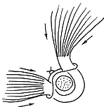
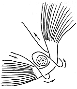

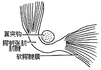
Figure 1: Method of tensor veli palatini tendon ligation.