| disease | Single Atrium |
Single atrium is a rare congenital heart disease caused by the failure of both the first and second septa of the atrial septum to develop during the embryonic period. There are no traces of the atrial septum, while the ventricular septum remains intact, hence it is also known as a two-ventricle three-chamber heart or single atrium three-chamber heart. Single atrium can exist alone but is often associated with a persistent left superior vena cava and dextrocardia, levocardia, or situs inversus of abdominal viscera. It is particularly common to find a cleft in the anterior leaflet of the mitral valve, and sometimes even atrioventricular canal defects.
bubble_chart Pathogenesis
The arterial and venous blood from the vena cava and pulmonary veins mixes in the single atrium. Due to the low resistance to filling in the right ventricle, most of the blood enters the left ventricle. Only a portion of the blood returning from the pulmonary veins to the atrium passes through the mitral valve into the left ventricle and then into the systemic circulation, which can clinically result in cyanosis. The oxygen saturation in the blood on both sides of the atrium, the ventricles, the main stirred pulse, and the pulmonary stirred pulse is almost the same. Single atrium combined with ectopic drainage of the vena cava is relatively common, such as the left superior vena cava draining into the coronary sinus or the left side of the common atrium. Next is the inferior vena cava Maijing azygos or hemiazygos vein drainage and hepatic veins directly entering the right side of the common atrium, forming mixed blood within the atrium.
bubble_chart Clinical Manifestations
The symptoms and signs are similar to those of a large atrial septal defect and atrioventricular canal defect. Common manifestations include crying, seasonal epidemics, acute cyanosis. Early onset of heart failure, gradually developing cyanosis and clubbing of fingers and toes, with an ejection murmur in the pulmonary valve area, a loud and fixed split second heart sound, and a systolic murmur of mitral insufficiency in the apical area.
bubble_chart Auxiliary Examination
Chest X-ray: Shows increased pulmonary vascular markings, enlarged cardiac silhouette, predominantly with right atrial and right ventricular enlargement, and a bulging pulmonary stirred pulse segment.
Electrocardiogram: Shows left axis deviation, frequent atrioventricular junctional rhythms, generally similar to atrioventricular canal defects.Cross-sectional echocardiogram: The echo reflection of the interatrial septum between the left and right atria is absent. In the four-chamber view, the normal cross-shaped echo reflection formed by the interatrial septum, interventricular septum, mitral valve, and tricuspid valve is altered to a T-shaped echo reflection.
Right heart catheterization: The catheter easily passes from the right atrium into the left atrium, or the catheter pathway resembles that of an atrioventricular canal defect. In fact, the single atrium contains mixed blood, so the oxygen saturation in the atria, ventricles, and the two major stirred pulses is roughly similar.
Selective atrial angiography: Can reveal a single atrium, and left ventricular angiography can show mitral regurgitation.
bubble_chart Treatment Measures
Surgical Indications: Single atrium, due to the presence of mixed blood within the atrium, can cause hypoxia and cyanosis, and may lead to cerebral embolism, infections, etc., due to increased red blood cells. Additionally, increased pulmonary circulation blood flow gradually leads to pulmonary stirred pulse hypertension, eventually forming irreversible pulmonary vascular obstructive sexually transmitted disease changes. Therefore, children with a clear diagnosis should strive for early surgery as long as severe pulmonary vascular obstructive sexually transmitted disease changes have not yet occurred.
The surgery is performed through a median sternotomy incision under extracorporeal circulation. A pericardial or Dacron patch is used to create an atrial septum. Those with left superior vena cava draining into the left atrium require simultaneous correction, and mitral valve anterior leaflet cleft with insufficiency requires repair.
(1) Repair of atrial septal defect: The atrial septal defect is repaired through a right atrial incision of the common atrium. The patch can be made of autologous pericardium, Dacron, or polytetrafluoroethylene artificial fabric, with superficial interrupted mattress sutures applied at the atrioventricular valve annulus. When suturing in the area of the atrioventricular node and bundle, it is best to keep the heart beating to prevent injury to the conduction system.
(2) Correction of left superior vena cava draining into the left atrium1. Simple ligation of the left superior vena cava: Applicable when there is an innominate vein communication between the left and right superior vena cava or sufficient collateral vessels.
2. Correction of left superior vena cava drainage into the right atrium: When there is no innominate vein between the bilateral superior vena cava and insufficient collateral vessels, the surgical method varies depending on the entry position of the left superior vena cava into the left atrium.
(1) Left superior vena cava entering at the upper left corner of the left atrium: Can be corrected simultaneously with the repair of the defect using a patch inside the atrium, suturing the upper end of the patch to the lower left edge of the left superior vena cava entrance, allowing venous return to directly enter the right side of the patch (Figure 1).
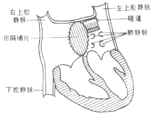
Figure 1 Schematic diagram of intracardiac patch correction surgery
(2) Left superior vena cava entering between the left pulmonary vein and the left atrial appendage: ① Rastelli method: The left superior vena cava internal drainage catheter is placed transversely on the posterior wall of the common atrial cavity, and the full thickness of the atrial posterior wall is wrapped around the catheter, sutured to form a tube. After removing the catheter, it becomes an intra-atrial tunnel, with the opening on the right side of the atrial septum, allowing blood flow from the left superior vena cava to enter the right atrium through this tunnel. ② Left superior vena cava entrance translocation: The left superior vena cava is cut from the left atrial entrance and anastomosed to the right atrial appendage (Figure 2). ③ Artificial vascular connection between the left superior vena cava and the right atrium.

Figure 2 Schematic diagram of left superior vena cava entrance transplantation
Single atrium needs to be differentiated from ventricular septal defect, total anomalous pulmonary venous return, complete transposition of the great arteries, tricuspid atresia, and complete atrioventricular canal defect. The clinical symptoms and signs of single atrium are similar to those of a large atrial septal defect or atrioventricular canal defect, but the symptoms appear earlier and are more severe. Cyanosis is present with increased pulmonary blood flow, and a large left-to-right shunt at the atrial level without significant evidence of pulmonary hypertension is characteristic.