| disease | Leukopenia and Agranulocytosis |
| alias | Leukopenia, Neutropenia, Agranulocytosis |
A peripheral blood white blood cell count below 4,000/mm3 is called leukopenia. Leukopenia is most commonly caused by neutropenia. An absolute neutrophil count below 1,800–2,000/mm3 is called neutropenia; below 500–1,000/mm3 is called agranulocytosis, often accompanied by severe and uncontrollable infections. The disease causes and mechanisms of disease for these three conditions are generally similar, but the severity of the illness varies.
bubble_chart Etiology
According to granulocyte kinetics and pathophysiology, this condition can be divided into four major categories:
(1) Bone marrow injury leading to reduced neutrophil production. Normally, adults generate a large number of neutrophils in the bone marrow daily, approximately 1011 or more. Colony-stimulating factors IL-3 and GM-CSF promote hematopoietic stem cells in the G0 phase to enter the cell proliferation cycle, while G-CSF specifically induces further proliferation and differentiation of granulocyte progenitor cells in the bone marrow proliferation pool. Metamyelocytes cease proliferation and continue to differentiate into banded and segmented neutrophils. These cells can remain in the bone marrow storage pool for about 5 days before entering the peripheral blood. Certain disease-causing factors directly injure the bone marrow, leading to quantitative or qualitative abnormalities in CFU-GM or hematopoietic dysfunction, which is often the most common cause of neutropenia:
1. Drug-induced injury. Antineoplastic drugs and immunosuppressants can directly kill proliferating cell populations. Drugs inhibit or interfere with granulocyte nucleic acid synthesis, affect cell metabolism, and hinder cell division. The direct toxic effects of drugs cause granulocytopenia, which is dose-dependent. Other classes of drugs may also have direct cytotoxic effects or reduce granulocyte production through immune mechanisms. (Table 1)
Table 1: Common drugs that can cause neutropenia
Category | Drug |
| Antineoplastic drugs | Nitrogen mustard, busulfan, cyclophosphamide, methotrexate, cytarabine, fluorouracil, doxorubicin, mitomycin, hydroxyurea, etc. |
| Anti-infective drugs | |
| Antibiotics | Penicillin G and other β-lactams, chloramphenicol, metronidazole |
| Sulfonamides | Compound formula sulfamethoxazole, sulfasalazine, sulfadiazine |
| Antituberculosis drugs | Isoniazid, isoniazone, rifampin, thioacetazone, para-aminosalicylic acid |
| Antimalarial drugs | Quinine, primaquine, chloroquine, pyrimethamine |
| Immunosuppressants | Azathioprine |
| Antiarrhythmic drugs | Procainamide, propranolol, quinidine |
| Antihypertensive drugs | Reserpine, hydralazine, methyldopa, captopril |
| Antithyroid drugs | Methylthiouracil, propylthiouracil, thiamazole, methimazole |
| Antipyretic and analgesic drugs | Aminopyrine, metamizole |
| Antirheumatic drugs | Phenylbutazone, indomethacin, ibuprofen, gold salts, penicillamine |
| Anticonvulsant drugs | Phenytoin sodium, dimethadione, phenobarbital, carbamazepine |
| Antipsychotic drugs | Wintermin, tricyclic antidepressants |
| Antidiabetic drugs | Tolbutamide (D860) |
| Diuretics | Mercury compounds, hydrochlorothiazide, ethacrynic acid |
| Others | Levamisole, cimetidine, metoclopramide, α and γ interferon, allopurinol |
2. Chemical toxins and radiation Chemical substances such as benzene and its derivatives, dinitrophenol, arsenic, bismuth, etc., have toxic effects on hematopoietic stem cells. X-rays, γ-rays, and neutrons can directly injure hematopoietic stem cells and the bone marrow microenvironment, causing acute or chronic radiation damage and resulting in granulocytopenia.
3. Immune factors Autoimmune granulocytopenia occurs when autoantibodies, T lymphocytes, or natural killer cells act on different stages of granulocyte differentiation, leading to bone marrow injury and impaired granulocyte production. This is commonly seen in Bi disease and autoimmune disorders. Certain drugs act as haptens in sensitive individuals, binding to granulocyte membrane proteins or plasma proteins to form complete antigens that adsorb onto granulocyte surfaces. These complete antigens stimulate the body to produce corresponding anti-granulocyte antibodies (IgG or IgM). Repeated drug use can cause granulocyte agglutination and destruction, known as immune drug-induced agranulocytosis. Some patients develop allergic reactions to certain drugs (sulfonamides, antipyretics, antibiotics, etc.), leading not only to granulocytopenia but also often accompanied by allergic manifestations such as rash, urticaria, asthma, and edema. Immune-mediated granulocytopenia is unrelated to drug dose.
4. Systemic infections Bacterial infections such as mycobacteria (especially subcutaneous nodule bacilli) and viral infections such as hepatitis viruses.
5. Abnormal cell infiltration of the bone marrow Cancer metastasis to the bone marrow, malignant hematopoietic system sexually transmitted diseases, and myelofibrosis can lead to bone marrow hematopoietic failure.
6. Impaired cell maturation—ineffective hematopoiesis For example, folate and vitamin B12 deficiency affects DNA synthesis. Bone marrow hematopoiesis is active, but cell maturation is arrested and destroyed within the bone marrow. Certain congenital granulocytopenia, acute non-lymphocytic leukemia, myelodysplastic syndromes, and paroxysmal nocturnal hemoglobinuria also exhibit maturation disorders, resulting in granulocytopenia.
(II) Abnormal distribution of peripheral circulating granulocytes Only half of the neutrophils entering the blood vessels are in the circulating pool, i.e., circulating with the blood flow, while the other half adhere tightly to the endothelial cells of capillaries and post-capillary venules (marginal pool) and do not circulate with the blood flow, thus not being detected in white blood cell counts. Granulocytes can shift between the circulating pool and the marginal pool. Injection of adrenaline or stress can rapidly transfer granulocytes from the marginal pool to the circulating pool, significantly increasing granulocyte counts. If the granulocyte count in the marginal pool increases relatively, it can cause pseudogranulocytopenia, where granulocyte production and utilization are normal. Systemic infections and allergic reactions can induce reactive acquired pseudogranulocytopenia (Figure 1).
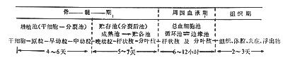
Figure 1 Schematic diagram of granulocyte kinetics
*Using 3H-pyrimidine in vitro DF32P measurement, the pool size is 6.5×13×109/kg, with a turnover time of 4 to 8 days. In vivo DF32P measurement shows the pool size to be 3~23×109/kg, with a turnover time of 8 to 14 days.
(3) Increased demand and accelerated consumption of granulocytes in extravascular tissues. Granulocytes typically remain in the blood vessels for only a few hours (with a half-life of 6 hours) before migrating to extravascular tissues to perform their defensive and waste-clearing functions, where they die within about 1–2 days. During bacterial, fungal, viral, or rickettsial infections, as well as allergic reactions, the production of granulocytes increases under the regulation of granulocyte-stimulating factors GM-CSF and G-CSF. This leads to more granulocytes being released from the bone marrow into the peripheral blood and tissues, along with enhanced phagocytic activity and bactericidal function. However, in severe infections, the body may fail to mount an adequate response to normal humoral stimuli. Concurrently, inflammatory mediators activate leukocyte adhesion molecules (such as CD11/CD18) on neutrophils and adhesion molecules (like ICAM-1) on vascular endothelial cells, facilitating leukocyte adhesion to vessel walls and migration across endothelial cells into tissues. Ultimately, transient granulocytopenia may still be observed in the blood. In autoimmune granulocytopenia and hypersplenism, granulocyte consumption may exceed the bone marrow's production capacity, leading to granulocytopenia.
(4) Mixed factors. Examples include chronic idiopathic granulocytopenia and cyclic granulocytopenia. Clinically, the three aforementioned types of leukopenia often coexist, necessitating careful analysis.
bubble_chart Clinical Manifestations
Leukopenia is often secondary to various systemic diseases, with clinical manifestations primarily dominated by the underlying condition. Most cases of leukopenia are transient and self-limiting, with no obvious clinical symptoms or nonspecific manifestations such as dizziness, lack of strength, low-grade fever, and pharyngitis. Neutrophils serve as the first line of defense against infections in the human body; thus, the clinical symptoms of granulocytopenia mainly involve susceptibility to recurrent infections. The risk of infection in patients is directly related to the neutrophil count, the duration of the decrease, and the rate of decline. Agranulocytosis (granulocytes <500/mm3) presents entirely differently from general leukopenia, with an acute onset. Due to the rapid destruction of a large number of granulocytes in a short period, patients may suddenly experience fear of cold, high fever, sweating, and general malaise. Severe infections almost invariably occur within 2–3 days. The lungs, urinary tract, oropharynx, and skin are the most common sites of infection. The mucous membrane may develop necrotic ulcers. Because of the lack of granulocytes mediating inflammatory responses, signs and symptoms of infection are often subtle; for example, severe pneumonia may show only mild infiltrates on chest X-rays without purulent sputum, severe skin infections may not form boils, and pyelonephritis may not present with pyuria. Infections can spread rapidly, progressing to sepsis, with a high mortality rate. The most common disease cause of this condition is drug reactions, and a relevant medical history may be present. Discontinuing the offending drug and receiving emergency treatment, followed by normalization of body temperature and recovery of peripheral white blood cell counts, indicates improvement.
Chronic idiopathic neutropenia is the most common type of chronic neutropenia. It is more prevalent in young and middle-aged women, with no clear history of specific medication use or exposure to chemicals. Clinically, patients may be asymptomatic or experience fatigue, low-grade fever, night sweating, or insomnia. Peripheral blood tests and bone marrow smears show no specific abnormalities.Cyclic neutropenia is characterized by recurrent periodic neutropenia accompanied by systemic lack of strength, fever, and grade I infections. Most patients develop symptoms in infancy, though onset may occur later. Several family members may be affected. The stage of attack lasts about 4–14 days, with an intermittent period of 12–35 days, during which symptoms may completely resolve.
Familial benign neutropenia is an autosomal dominant genetic disorder with a later onset, intermittent episodes, and grade II granulocytopenia, with a generally favorable course. Bone marrow examination shows granulocyte maturation arrest at the myelocyte and metamyelocyte stages, possibly accompanied by hypogammaglobulinemia. Symptoms may spontaneously resolve with age.
Patients with only neutropenia, no underlying disease, and no recurrent infections may be collectively referred to as having benign neutropenia, including familial, congenital, and pseudoneutropenia.
The white blood cell count is the primary diagnostic laboratory criterion. The white blood cell count can fluctuate significantly due to various influencing factors, so repeated testing is often necessary for diagnosis. Toxic granules and vacuoles may be present in the cytoplasm of granulocytes, often indicating a bacterial infection. The proportion of monocytes is frequently compensatorily increased. If the proportion of band forms increases (>20%), it suggests that the bone marrow has sufficient granulocyte production capacity. The bone marrow findings vary depending on the primary disease. In agranulocytosis, the bone marrow shows a severe reduction or even complete absence of neutrophils at all stages. Granulocytes may exhibit marked toxic changes or maturation arrest. Lymphocytes, monocytes, plasma cells, and histiocytes may increase, while erythroblasts and megakaryocytes are generally normal. During recovery, late-stage myelocytes and more mature granulocytes appear sequentially in the peripheral blood, and in some cases, a leukemoid blood picture may be observed.
The second step in diagnosis is identifying the disease cause of leukopenia. It is important to inquire about potential exposure to drugs or chemicals that may induce the condition, underlying diseases that cause granulocytopenia (e.g., chronic inflammation, autoimmune diseases), and a history of recurrent infections. The following special tests can assist in understanding the disease cause and pathogenesis of granulocytopenia.
(1) Measurement of bone marrow granulocyte reserve function: Using pyrogens such as etiocholanolone, lipopolysaccharide, or prednisone (or hydrocortisone) to stimulate the release of bone marrow granulocytes through the action of an intermediate product—"neutrophil-releasing factor"—to assess granulocyte release function.
If the white blood cell count increases by more than 2×10⁹/L five hours after oral administration of 40mg prednisone or by more than 4×10⁹~5×10⁹/L 3–4 hours after intravenous injection of 200mg hydrocortisone, the result is considered normal.
(2) Epinephrine test: After subcutaneous injection of 0.2mg, the white blood cell count is measured after 20 minutes. If it increases by 2000/mm3 or more than doubles the baseline level, it suggests excessive granulocyte aggregation in the marginal pool of blood vessel walls. If there is no splenomegaly, pseudogranulocytopenia may be considered.
(3) Leukocyte agglutinins: In some patients with immune-mediated granulocytopenia, leukocyte agglutinins may be detected in the serum, which has auxiliary diagnostic significance. However, individuals who have received multiple blood transfusions or multiparous women may also test positive.
(4) Lysozyme: Measuring serum and bone marrow lysozyme levels can help assess granulocyte production.
bubble_chart Treatment Measures
Patients with secondary granulocytopenia should actively treat the underlying disease and discontinue exposure to suspected drugs or toxins. Treatment methods should be selected based on different pathological mechanisms.
(1) Drug Therapy Lithium carbonate can increase granulocyte production but is ineffective for chronic bone marrow failure. The adult dose is 300mg, taken orally three times daily. After effectiveness is observed, the dose should be reduced to 200mg twice daily for maintenance over 2–4 weeks. Side effects may include tremor, gastric discomfort, diarrhea, cutaneous pruritus, edema, etc., which disappear upon discontinuation. Use with caution in patients with kidney disease. Adrenocortical hormones or azathioprine may be effective for immune-mediated granulocytopenia. Long-term follow-up with stable blood counts and no infection generally does not require medication.
(2) Recombinant Human Granulocyte Growth Factors GM-CSF and G-CSF can induce hematopoietic stem cells into the proliferation cycle, promote granulocyte proliferation, differentiation, and maturation, and release them from the bone marrow into peripheral blood. They also enhance granulocyte chemotaxis, phagocytosis, and bactericidal activity. G-CSF is more effective for cyclic granulocytopenia and severe congenital agranulocytosis in children. It accelerates the recovery of chemotherapy-induced leukopenia and can also be used to prevent leukopenia and fever caused by intensive chemotherapy. Depending on the condition, administer 50μg/m2 subcutaneously once daily or 100–300μg/d via subcutaneous or intravenous infusion. Adjust or discontinue the dose as appropriate after white blood cell recovery. Side effects of CSF include fever, shivering, bone arthralgia, etc.
(3) Anti-infective Therapy Once a patient develops fever, blood, urine, and other relevant cultures should be performed, and broad-spectrum antibiotics should be administered immediately. Switch to targeted agents after the pathogen is identified. If the pathogen cannot be confirmed, empirical treatment with broad-spectrum antibiotics must be completed in full course, with attention to preventing secondary infections such as fungi or anaerobes. Patients with acute agranulocytosis must receive strict disinfection and isolation protection, preferably in an air-purified sterile room, with enhanced skin and oral care to prevent cross-infection. Anti-infective therapy is often critical to the success of rescue efforts in agranulocytosis.
(4) Others Transfusion of concentrated granulocyte suspensions has been attempted for severe infections but is rarely used now due to the rapid production of granulocyte antibodies in recipients. For cases where bone marrow failure is the primary cause of agranulocytosis and severe symptoms are not mediated by immunity, allogeneic hematopoietic stem cell transplantation may be considered.